
Characterization of Pictorial Materials of Two 18 Century Sculptures: King David and Bathsheba
Sanches, F.A.C.R.A., Nardes, R.C., dos Santos, R.S.; Leitão, R.G.; Leitão, C.C.G.; Assis, J.T.; de Gusmão, E.T.; Lopes, R.T.; de Oliveira, D.F.; dos Anjos, M.J.
DOI 10.5433/1679-0375.2023.v44.47972
Citation Semin., Ciênc. Exatas Tecnol. 2023, v. 44: e47972
Abstract:
In this work, the X-ray fluorescence technique was employed to characterize the pictorial materials used in two sculptures from the 18\(^{ ext{th}}\) century. In addition, Raman spectroscopy was used to characterize the preparation layers used in the sculptures. The analyzed sculptures are carved in wood, with gilding and polychrome, and represent the biblical characters, Bathsheba, and King David. The sculptures are property of the Church of extitNossa Senhora do Pilar, in Duque de Caxias, RJ. XRF analyzes were performed using a portable ED-XRF system, which has a low-power X-ray tube (Amptek) with a silver target (Ag) and an SDD detector (Amptek). Raman spectroscopy analyzes were performed using the DXR2 Raman microscopy equipment (Thermo Fisher Scientific) with a 785 nm laser source. The results obtained from the XRF and Raman spectroscopy techniques suggest the presence of Gypsum, Calcite, Calcium Sulfate. In addition, the results obtained suggest the presence of the following pigments in the sculptures: Lead white, Titanium White, Lithopone, Ochre, Vermilion, Red Lead and gilding with gold leaf. The studies also showed that the Bathsheba sculpture was probably subjected to some processes of chromatic reintegration over the years due to the heterogeneity of pigments found in the same region. space-0.1cm
Keywords: archaeometry, X-ray fluorescence, raman spectroscopy, baroque sculpture space-0.1cm
Introduction
The use of non-destructive techniques has been decisive in the scientific analysis of cultural heritage and works of art. Due to the unique features and historical importance of cultural heritage objects, the research must be carefully planned to preserve the analyzed artifact and obtain as much information as possible about the piece.
Scientific investigations can be conducted using multiple non-destructive or minimally invasive analytical techniques. In some cases, an important factor in choosing the techniques used is their portability, as many works of art cannot be removed from the museum, requiring an on-site analysis (Vandenabeele & Donais, 2016).
Information about the chemical composition of artworks and cultural heritage is important for the better conservation and preservation of the artifact since certain materials can be sensitive to light, moisture, or damp conditions, heat, and air pollutants (Spring & Gorout, 2002; Ali & Mansour, 2018; Bonizzoni et al., 2018).
Characterization of the pigments helps to understand the history of that period, the technologies used in the manufacture, and whether the artifact has already gone through some intervention process (Adriaens, 2005; Sanches et al., 2022).
X-ray fluorescence (XRF) and Raman spectroscopy are useful tools for characterizing historic pigments. The associated use of these techniques has been confirmed by numerous authors, and successful field applications have been reported in Felix et al., 2018, Freitas et al., 2016, Ricci et al., 2004, Sanches et al., 2022, and Sawczak et al., 2009.
XRF provides information about the elemental composition of the material. The characterization of pictorial materials by XRF is identified by the presence of key elements in the XRF spectra associated with the color of the analyzed region. However, not all elements detected in the XRF spectra are characteristic of the pigment used. Commonly appears contributions of XRF spectral lines come from the preparation layers, from the overlapping of pigments, from support (wood) and from trace elements or contaminants, whose presence does not contribute to the optical properties of the pigment (Clark, 2002; Zuena et al., 2021).
Raman spectroscopy offers molecular information about the material analyzed. The technique allows the identification of organic and inorganic compounds, such as crystalline and amorphous structures, by analyzing a small amount of samples (Tomasini et al., 2012; Coccato et al., 2015).
In this work, the X-ray fluorescence technique was employed to characterize the pictorial materials used in two sculptures from the 18\(^{\text{th}}\) century. In addition, spectroscopy Raman was used to characterize the preparation layers used in the sculptures.
Materials and methods
Analyzed artifacts
The two sculptures analyzed represent the biblical characters of King David and Bathsheba and belong to the Mother Church of Nossa Senhora do Pilar, located in Duque de Caxias, Rio de Janeiro, Brazil. The church was built in 1720 and was listed by the National Heritage in 1938 (Rodrigues & Mello, 2020).
According the to historian Fabrino, 2012, the Church of Nossa Senhora do Pilar was the target of many criminal actions over the years, and many of its sacred art pieces were stolen. Including the sculptures of King David and Bathsheba, which were stolen in 1974 and remained missing for four decades. The sculptures were recovered thanks to the efforts of the Diocese of Duque de Caxias together with the Brazilian competent authorities (Alves, 2021).
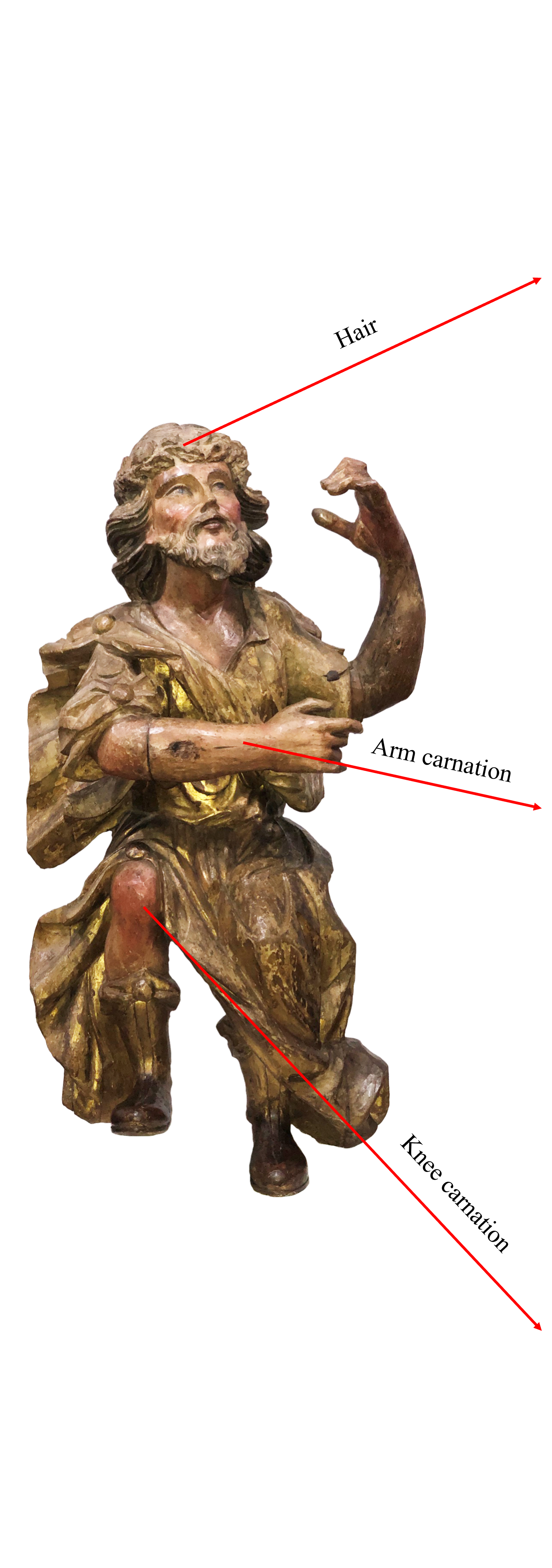
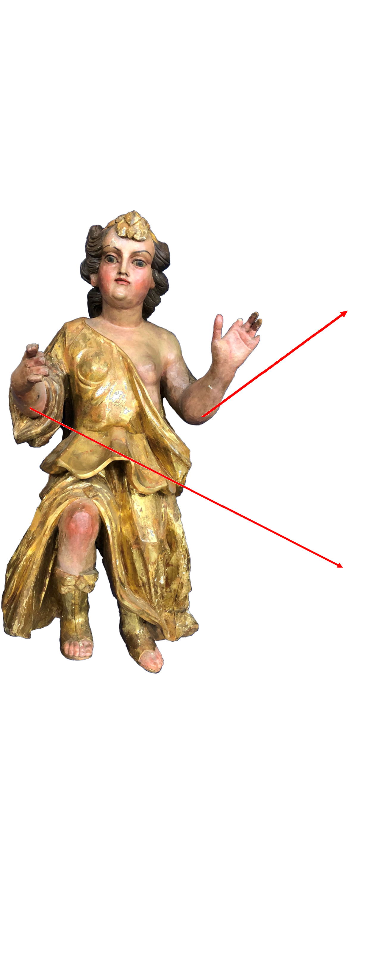
The two analyzed pieces are sculptures carved in wood, gilded, and polychrome. Figure 1 shows images of King David and Bathsheba. Both sculptures are approximately 1.20 m long. King David is approximately 75 cm wide and 40 cm deep, and Bathsheba is approximately 50 cm wide and 55 cm deep.
| (a) | (b) | |
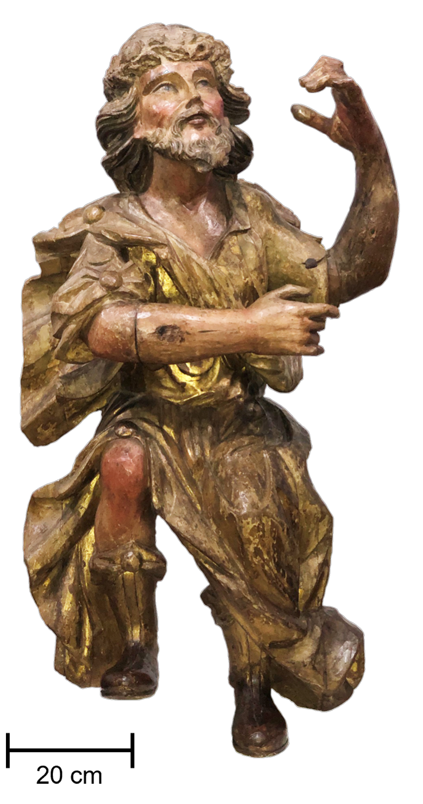 | 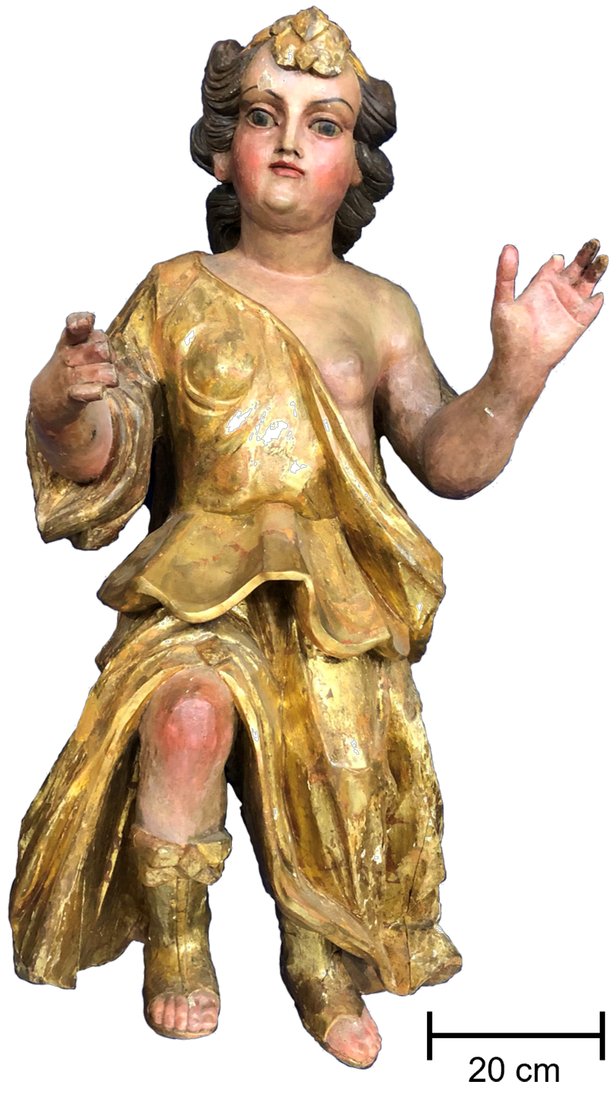 |
The author of the sculptures of Figures 1(a) and 1(b) is unknown, as is the date of execution. It is only known that they were executed in the first half of the 18\(^{\text{th}}\) century, with characteristics of the Joanine Baroque, in polychrome wood and gilding.
Experimental
The pigments and preparation layers of the statues of King David and Bathsheba were analyzed using a portable Energy Dispersion X-Ray Fluorescence (ED-XRF) system developed at the Laboratory of Electronic Instrumentation and Analytical Techniques (LIETA/IFADT/UERJ).
The portable ED-XRF system consists of a low-power (4 W) silver target X-ray tube model mini-X (Amptek) and a silicon drift detector model X-123 (Amptek) with an energy resolution of 122 eV at 5.9 keV. The measurements were performed under the following experimental conditions: voltage of 40 kV, current of 50 µA, during an acquisition time of 100 s. XRF measurements were distributed in the wood, preparation layers, carnation pigments, hair, and gilding in the two sculptures.
All XRF spectra were evaluated using the open-source Python Multichannel Analyzer (PyMCA) software package (Solé et al., 2007).
Samples from the preparation layer were analyzed ex-situ using the Raman spectroscopy technique, which was performed using Raman DXR2 - Thermo Scientific microscopy equipment (Thermo Fisher Scientific). A 785-nm laser source with a set power of 4 mW, and a focused 20x objective excited the samples. Spectra were collected over a range of 100-2000 cm\(^{-1}\), with an acquisition time of two seconds and four accumulations.
Results and discussion
King David
Figure 2 shows the XRF spectra of the wood and the preparation layer of the King David sculpture.
| (a) | (b) |
 |  |
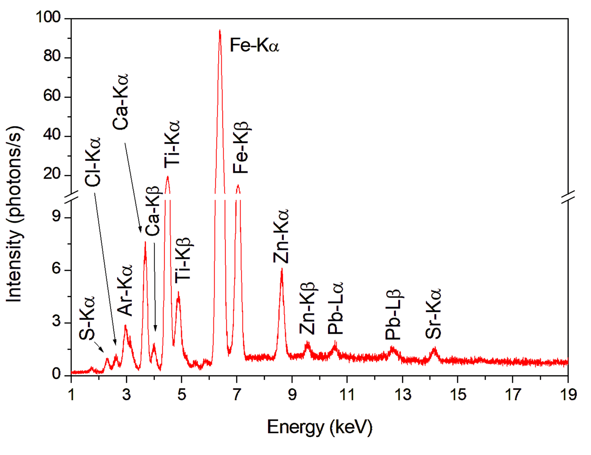
The first XRF measurement was performed in a region without pigment to identify the key elements related to wood, illustrated in Figure 2(a). The elements detected in the wood were S, K, Ca, Ti, Fe, Zn, Br, and Sr.
In the XRF analysis of the preparation layer, Figure 2(b), elements S, Ca, Fe, and Sr were detected. The elements S and Ca can be considered to be the key elements of the preparation layer. A complementary analysis with Raman spectroscopy was also performed, and the detected bands were: 416 cm\(^{-1}\), 495 cm\(^{-1}\) and 1010 cm\(^{-1}\), which are related to calcium sulfate dihydrate, suggesting that the preparation layer was made by gypsum (CaSO\(_4\)·2H\(_2\)O) (Prieto-Taboada et al., 2014).
The production of polychrome wood sculptures in Brazil during the Baroque period (18\(^{\text{th}}\) and 19\(^{\text{th}}\) centuries) has similar characteristics in the composition of the preparation layer. Before receiving the polychrome, the artist applied successive layers of plaster to correct imperfections in the carving and produce a smooth surface to receive the pictorial layer (Coelho, 2005).
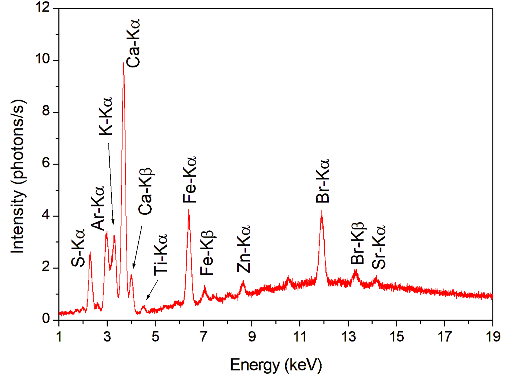
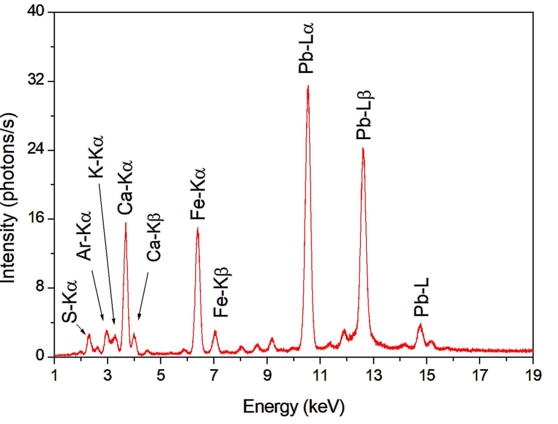
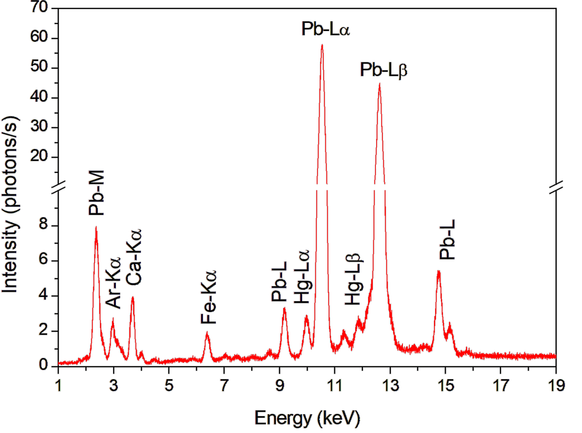
Figure 3 shows the XRF spectra from brown pigments (hair) and carnation areas (arm and knee) of the King David sculpture.
The XRF spectra of the brown pigment, Figure 3(a), showed the the following elements: S, Ca, Fe, and Pb. The element Fe can be identified as the key element for brown pigment, which suggests a mixture of an ocher pigment (Fe\(_2\)O\(_3\)) with carbon black (C), which is not detected by XRF, associated with lead white (2PbCO\(_3\).Pb(OH)\(_2\)) as an extender.
 | (a) |
 | |
| (b) | |
 | |
| (c) | |
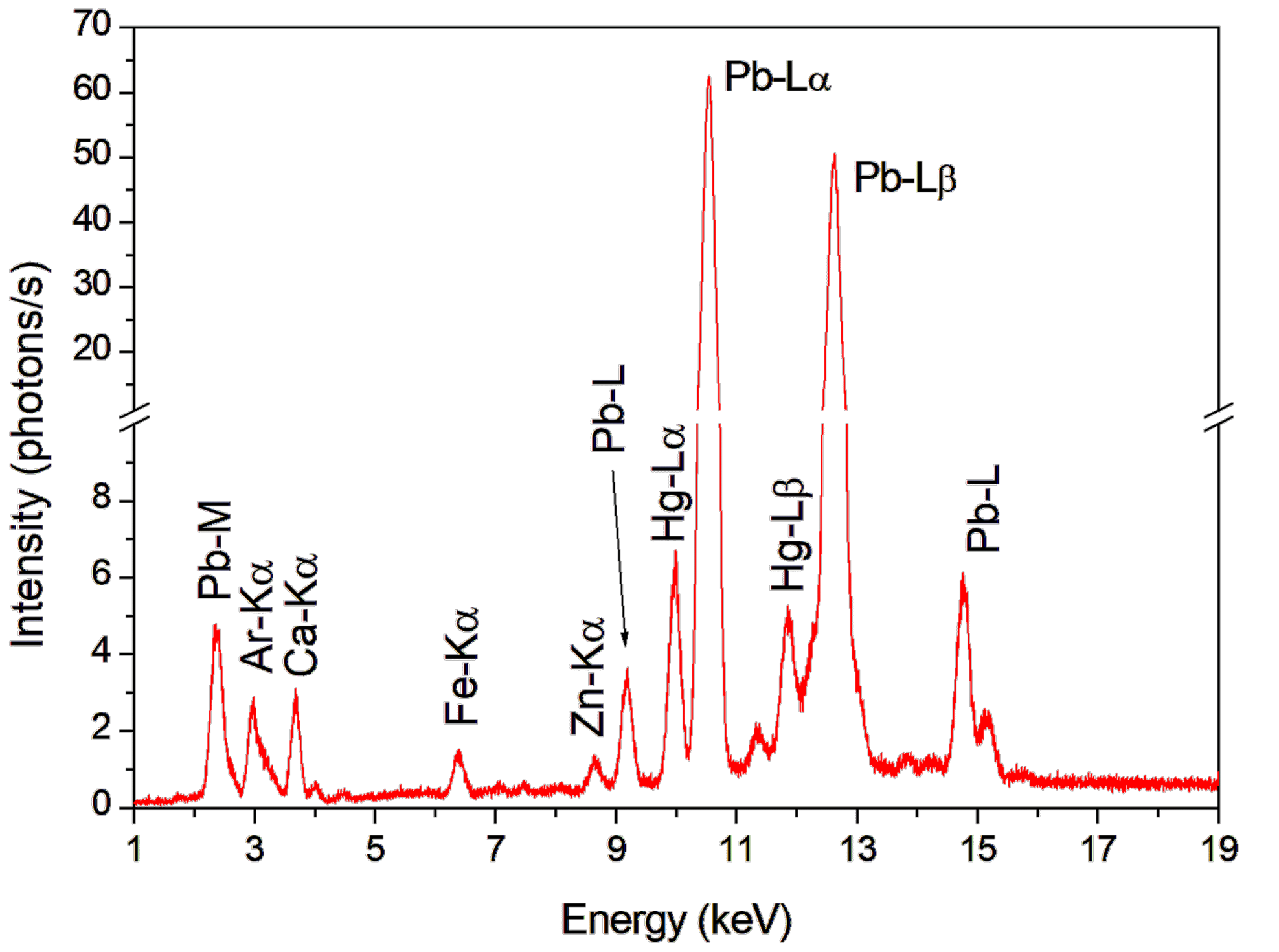 |
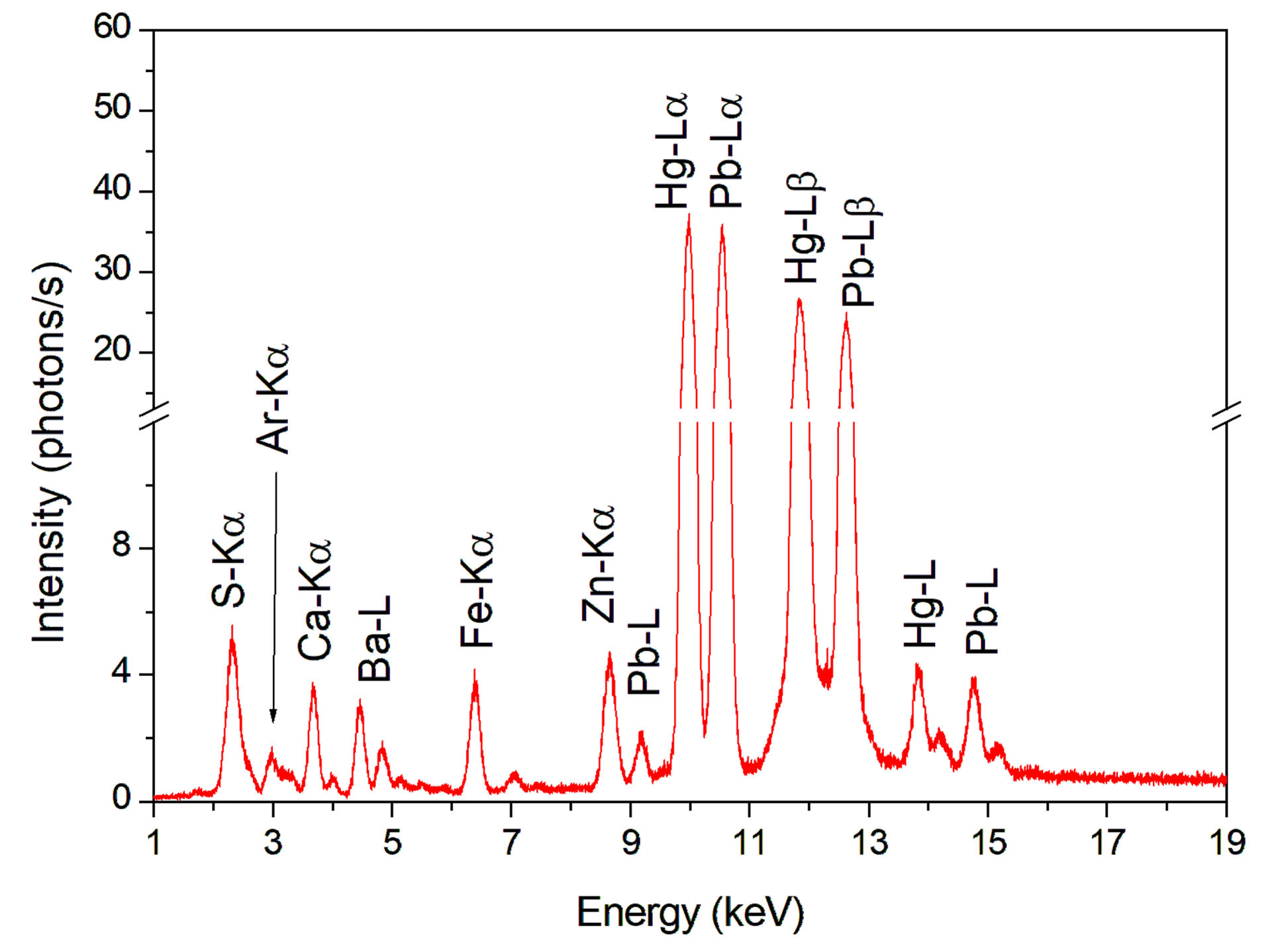
In the XRF carnation’s spectra, Figures 3(b) and 3(c), it was possible to detect the key elements Hg and Pb. The element Hg in this region of the carnation suggests the presence of pigment vermilion (HgS). Vermilion is a red pigment that has been used since antiquity, however its use was drastically reduced after the introduction of cadmium red (CdS) in 1919 (Spring & Gorout, 2002). The element Pb indicates the presence of lead white (2PbCO\(_3\).Pb(OH)\(_2\)) or minium (Pb3O4), that are sometimes used in mixtures (Bruni et al., 1999).
The presence of lead white suggests it was used as an extender for the red pigment. It is possible to notice the difference in the Hg intensity between the XRF spectra of Figures 3(b) and 3(c). In the carnation area where the red tone is lighter (arm), the Hg element intensity decreases.
Figure 4 shows the XRF spectrum of the gilding of the King David sculpture. The elements detected in the gold leaves, used especially on the clothing areas, were Ca, Ag, Fe, Cu, Au, Sr, Ba, and Pb.
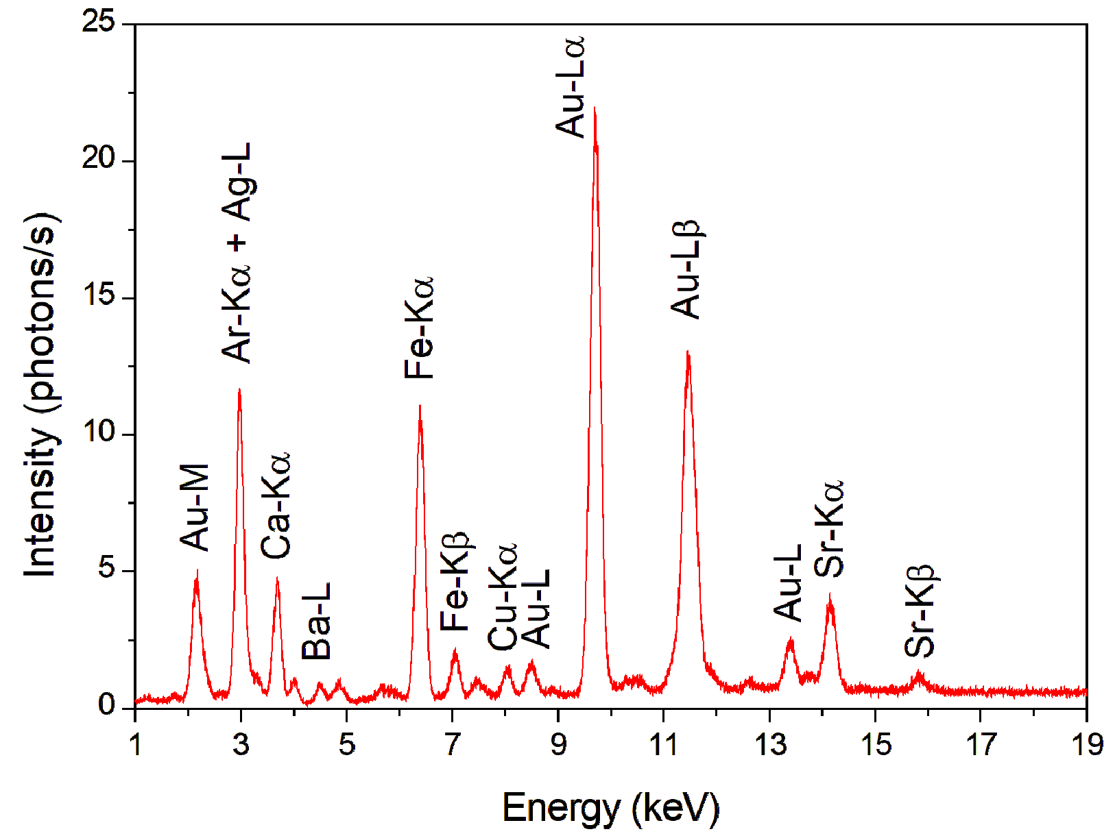
The high count of Fe, in Figure 4, suggests that the gilding technique used in the King David sculpture was water gilding, which uses an Armenian bole to fix the gold leaf to the support (Gulotta et al., 2012; Hradil et al., 2017).
Despite the X-ray tube target being silver, the increase in silver L-line counts suggests that silver is also present in the gold leaf alloy. Thus, the metallic alloy of Au sheets can be composed of Au and Ag (Le Gac et al., 2009).
Bathsheba
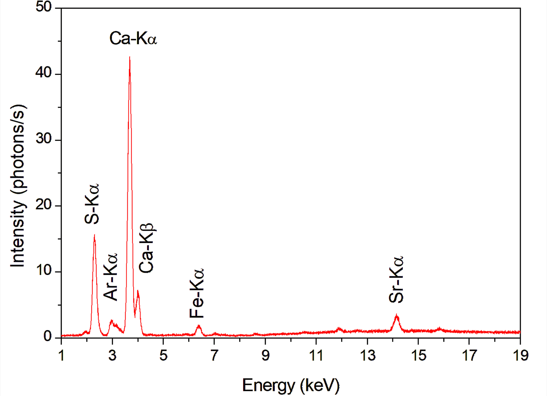
Figure 5 shows the XRF spectra of the wood and preparation layer of the Bathsheba sculpture. In the XRF spectrum of the wood was detected the elements Si, S, Ca, Ti, Fe, Br, Sr and Pb, Figure 5(a). While, in the XRF analysis of the preparation layer, Figure 5(b), it was detected the elements S, Ca, Fe, Br and Sr.
| (a) | (b) |
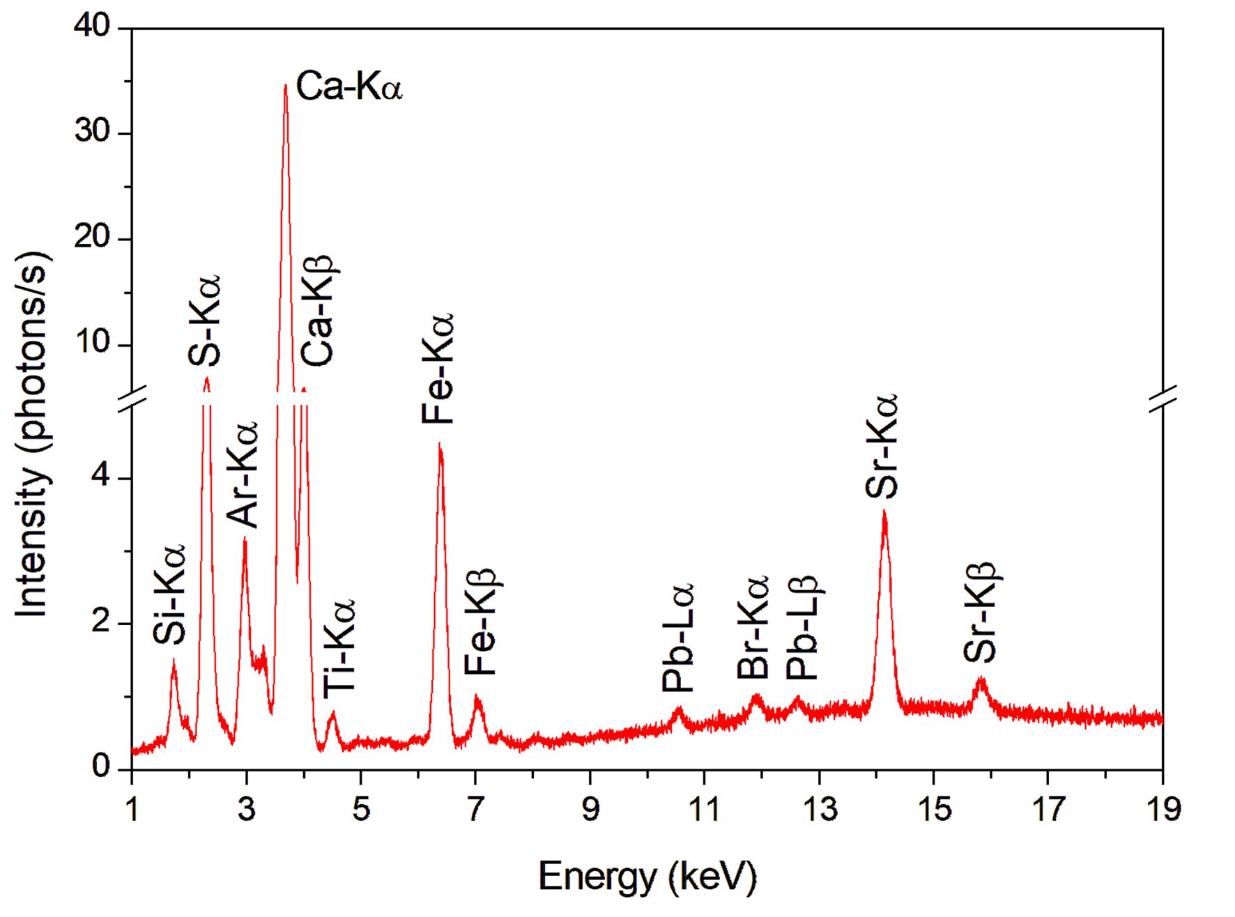 |  |
It is possible to notice that the XRF spectra of the wood and the preparation layer are similar. In the Bathsheba sculpture, it was not possible to perform XRF measurements of the wood on the back of the sculpture (region without the addition of pigments). The analyzes of the wood and preparation layer were carried out in regions of pigment loss.
The elements S and Ca can be considered the key elements of the preparation layer and indicate the presence of calcium sulfate (CaSO\(_4\)).
However, Raman spectroscopy analysis of the preparation layer detected the bands 156 cm\(^{-1}\), 281 cm\(^{-1}\), 712 cm\(^{-1}\) and 1086 cm\(^{-1}\), which are related to calcite (CaCO\(_3\)) (Kontoyannis & Vagenas, 2000). These results suggest that the preparation layer can contain both materials.
Figure 6 shows the XRF spectra of the brown pigments (hair), mouth and knee carnations.
| (a) | |
 | |
| (b) | |
 | |
| (c) | |
 |
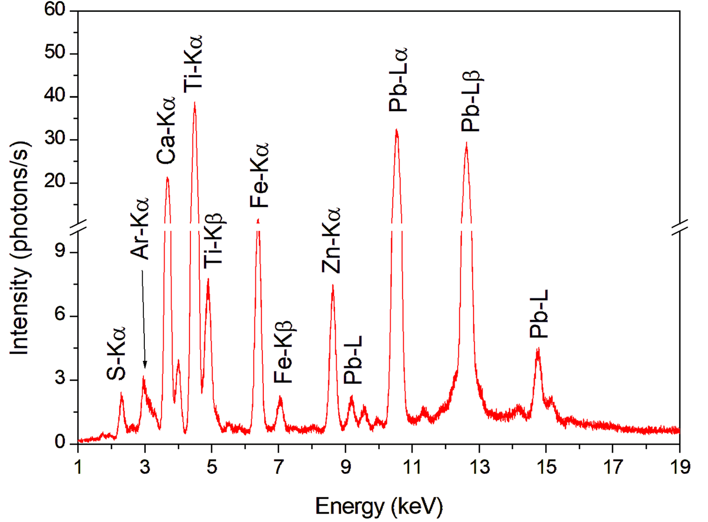
In the XRF spectrum of the brown pigment, Figure 6(a), Bathsheba’s head, elements S, Cl, Ca, Ti, Fe, Zn, Sr, and Pb were identified. The key element for this pigment is Fe, which is associated with earth pigments such as ocher (Fe\(_2\)O\(_3\)). The presence of Ti with high intensities indicates that the pigment used as an extender in this region could be titanium white (TiO\(_2\)).
Titanium white pigment became available for wide commercialization at the beginning of the 20\(^{th}\) century and is the most common white pigment today due to its bright color, opacity, and non-toxic character (Lauridsen et al., 2014). This suggests that there was a repainting process after the beginning of the 20\(^{th}\) century.
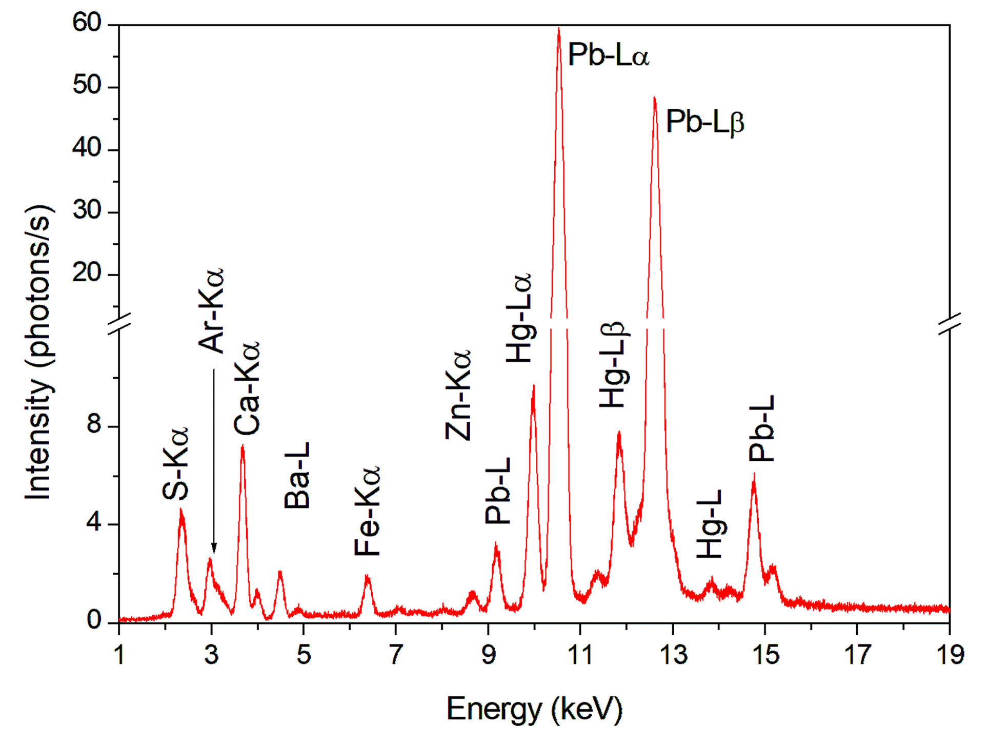
In the pigment analyzed in the region of the mouth and knee, Figures 6(b) and 6(c), it was possible to detect the elements S, Ca, Ba, Fe, Zn, Hg, and Pb. The key elements are Hg and Pb. The element Hg in this region of the carnation suggests the presence of the pigment vermilion (HgS). The presence of Pb suggests that the lead white pigment (2PbCO\(_3\).Pb(OH)\(_2\)) was used as an extender for the vermilion pig- ment.
According to the XRF spectra in Figures 6(b) and 6(c), it is possible to notice that the concentrations of these elements vary according to the chosen tone. The Pb count increases significantly in lighter tone areas, such as the knee of the Bathsheba sculpture.
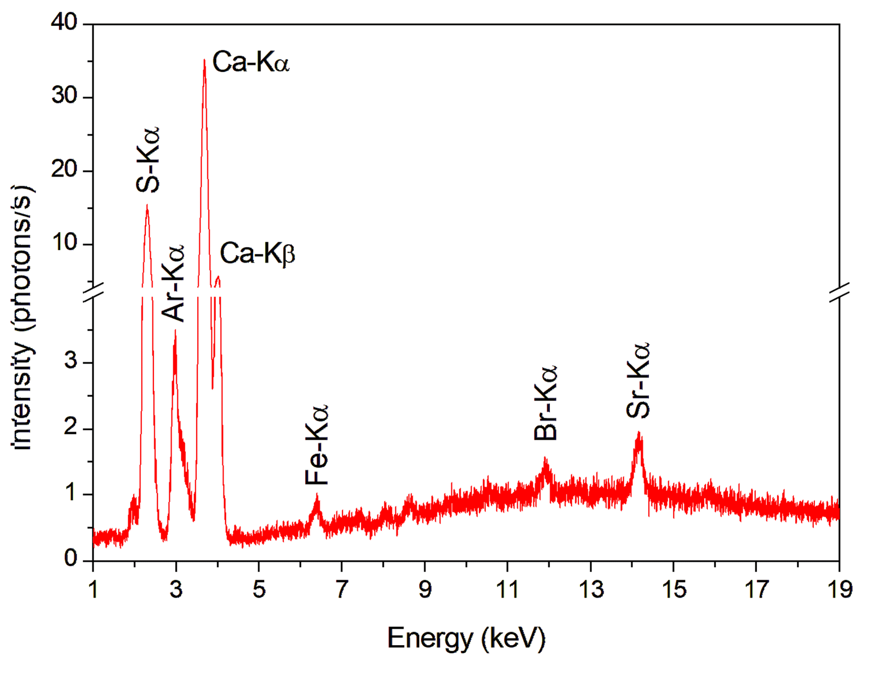
Figure 7 shows the right and left arms of the XRF spectrum of carnation.
In the carnation area (arms) of the Bathsheba sculpture, the XRF spectra showed different chemical compositions, Figure 7(a) and 7(b). This non-homogeneity of the pigment used in the carnation is an indication that the piece has undergone chromatic reintegration processes over the years.
 | (a) |
 | |
| (b) | |
|
In the XRF spectrum of the left arm carnation, Figure 7(a), the elements S, Ca, Ti, Fe, Zn, and Pb were identified. In this XRF spectrum, the element Ba was not detected. The presence of Pb suggests the presence of minium (Pb\(_3\)O\(_4\)), and the high intensities of Ti may indicate that the titanium white pigment was used as an extender in this region.
The elements S, Ca, Ba, Fe, Zn, and Pb were detected in the right arm carnation, Figure 7(b). The Hg element was not detected, and the high Pb count suggests the presence of red lead, also known as minium (Pb\(_3\)O\(_4\)).
Minium was used from antiquity until the 19\(^{th}\) century. Its use was common in carnation zones in Portuguese sculptures executed in wood from the Baroque period (Barata et al., 2013).
The red lead (Pb\(_3\)O\(_4\)) and lead white (2PbCO\(_3\).Pb(OH)\(_2\)) pigments can be responsible for the carnation color, both of which were discontinued in the 19\(^{th}\) century.
The presence of Ba and Zn elements suggests the lithopone (BaSO\(_4\).ZnS). This pigment was synthesized for the first time in 1850 and is a mixture of barium sulfate and zinc sulfide (Palamara et al., 2021).
Figure 8 shows the difference between the XRF spectra from the original and reintegration gilding. In the original gilding regions, it was possible to verify the presence of Ca, Ti, Fe, Zn, Sr, Ag, Au, and Pb elements.
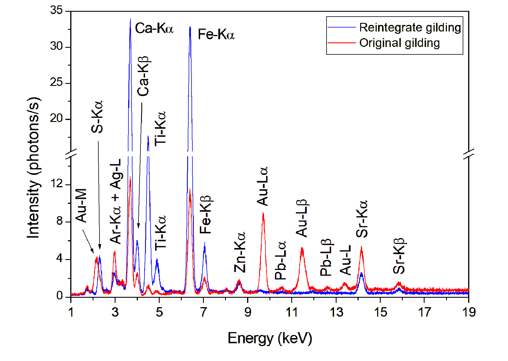
In the XRF spectrum of the gold leaf, it was possible to see high intensities of Ca, Fe, and Sr. These elements may be associated with the Armenian bole, which was found as to be a preparation base for the application of gold leaf, but the XRF technique was appropriate to characterize it. The presence of Ag suggests that the Au leaf is composed of a metallic alloy of Au and Ag, such as in the gilding of King David’s sculpture.
It was also possible to identify the regions of chromatic reintegration of the gilding. The elements found were S, Ca, Ti, Fe, Zn, and Sr. This chromatic reintegration may consist of a mixture of different colors to obtain a golden tone. Unfortunately, the XRF technique was unable to characterize the pigment.
Table 1 shows all the key elements found in each region analyzed in the King David and Bathsheba sculptures and the suggested pigments.
| Sculpture | Analyzed area | Key elements | Suggested pigments |
|---|---|---|---|
| King David | |||
| Wood | S, K, Ca, Ti, Fe, Zn, Br, Sr | - | |
| Preparation layer | S and Ca | Gypsum | |
| Brown (hair and beard) | Fe | Ocher | |
| Carnation | Hg and Pb | Vermilion and lead white | |
| Gilding | Ag and Au | Gold leaves | |
| Bathsheba | |||
| Wood | Si, S, Ca, Ti, Fe, Br, Sr, Pb | - | |
| Preparation layer | S and Ca | Calcite and calcium sulfate | |
| Brown (hair) | Fe and Ti | Ocher and titanium white | |
| Mouth and knee carnation | Hg and Pb | Vermilion and lead white | |
| Arms carnation | Ti, Zn, Ba, Pb | Red lead, lead white, lithopone, | |
| and titanium white | |||
| Original gilding | Ag and Au | Gold leaves | |
| Reintegrated gilding | S, Ca, Ti, Fe | - | |
Conclusions
Using the XRF technique and Raman spectroscopy, it was possible to characterize the pictorial materials and the preparation layer of the King David and Bathsheba sculptures.
In the King David sculpture, the results obtained suggest the presence of gypsum in the preparation layer, vermilion and lead white in the carnation areas, and ocher pigment in the hair and beard. In addition, the gold leaf used in the gilding of the sculpture of King David is composed of a metallic alloy of Ag and Au.
On the other hand, in the analysis of the Bathsheba sculpture, the results obtained suggest the presence of gypsum and calcite in the preparation layer. In the mouth and knee carnations, the suggested pigments were lead white and vermilion. However, the pigments red lead, lead white, lithopone, and titanium white are suggested for the arms carnation region. The analysis also showed that the Bathsheba sculpture was probably subjected to some processes of chromatic reintegration over the years due to the non-homogeneity of pigments in the same region and the presence of modern pigments such as titanium white.
The gold leaf used in the gilding of the Bathsheba sculpture is composed of a metallic alloy of Au and Ag. In addition, in the regions of loss of gold leaf, it was possible to identify chromatic reintegration.
Author contributions
F.A.C.R.A. Sanches, R.C. Nardes, R.S. dos Santos participated in the: Conceptualization, Data Curation, Formal Analysis, Investigation, Methodology, Writing. R.G. Leitão, C.C.G. Leitão, J.T. Assis, E.T. de Gusmão, R.T. Lopes, D.F. de Oliveira participated in the: Resources. M.J. Anjos participated in the: Projects Managements, Supervision, Validation, Visualization.
Conflicts of interest
The authors declare no conflict of interest.
Acknowledgments
This study was partially funded by Conselho Nacional de Desenvolvimento Científico e Tecnológico (CNPq), Fundação Carlos Chagas Filho de Amparo à Pesquisa do Estado do Rio de Janeiro (FAPERJ) and Coordenação de Aperfeiçoamento de Pessoal de Nível Superior - Brasil (CAPES) – code Financial 001 and Financiadora de Estudos e Projetos (FINEP - CT - INFRA 01.13.0444.02).
References
Adriaens, Annemie (2005). Non-destructive analysis and testing of museum objects: An overview of 5 years of research. 1503--1516. https://doi.org/10.1016/j.sab.2005.10.006
Ali, M. F. & Mansour, M. M. A. (2018). A study of biodeterioration and chromatic alterations of painted and gilded mummy cartonnage at the Saqqara Museum Storeroom, Egypt. 845--858. https://doi.org/10.1111/arcm.12340
Alves, M. (2021). Furtada há 47 anos, imagem de Rei Davi é recuperada pela Diocese de Duque de Caxias. Diário do Rio. https://diariodorio.com/furtadaha-47-anos-imagem-de-rei-davi-e-recuperadapela-diocese-de-duque-de-caxias/
Barata, C., Carballo, J., António João Cruz, A. J., Coroado, J., Araújo, M. E., & Mendonça, M. H. (2013). Characterization by chemical analysis of Portuguese baroque polychrome wooden sculptures with erudite and popular features. Química Nova, 36(1), 21–26. http://www.scopus.com/inward/record.url?eid=2-s2.0-84874486331&partnerID=MN8TOARS
Bonizzoni, L., Bruni, S., Gargano, M., Guglielmi, V., Zaffino, C., Pezzotta, A., Pilato, A., Auricchio, T., Delvaux, L. & Ludwig, N. (2018). Use of integrated non-invasive analyses for pigment characterization and indirect dating of old restorations on one Egyptian coffin of the XXI dynasty. 122--131. https://doi.org/10.1016/j.microc.2018.01.002
Bruni, S., Cariati, F., Casadio, F. & Toniolo, L. (1999). Identification of pigments on a XV century illuminated parchment by Raman and FTIR microspectroscopies. 55(7-8), 1371--1377. https://doi.org/10.1016/S1386-1425(98)00300-X
Clark, R. J. H. (2002). Pigment identification by spectroscopic means: an arts/science interface. 7--20. https://doi.org/10.1016/S1631-0748(02)01341-3
Coccato, A., Jehlicka, J., Moens, L. & Vandenabeele, P. (2015). Raman spectroscopy for the investigation of carbon-based black pigments. 46(10), 1003--1015. https://doi.org/10.1002/jrs.4715
Coelho, B. R. V. (2005). Materiais, técnicas e conservação. In B. C. Coelho (org), Devoção e arte: Imaginária religiosa em Minas Gerais (pp. 233–245). Universidade de São Paulo.
Felix, V. S., Mello, U. L., Pereira, M. O., Oliveira, A. L., Ferreira, D. S., Carvalho, C. S., Silva, F. L., Pimenta, A. R., Diniz, M. G. & Freitas, R. P. (2018). Analysis of a European cupboard by XRF, Raman and FT-IR. 198--204. https://doi.org/10.1016/j.radphyschem.2018.06.036
Freitas, R. P., Ribeiro, I. M., Calza, C., Oliveira, A. L., Felix, V. S., Ferreira, D. S., Pimenta, A. R., Pereira, R. V., Pereira, M. O. & Lopes, R. T. (2016). Analysis of a Brazilian baroque sculpture using Raman spectroscopy and FT-IR. 67--71. https://doi.org/10.1016/j.saa.2015.10.013
Gulotta, D., Goidanich, S., Bertoldi, M., Bortolotto, S. & Toniolo, L. (2012). Gildings and false gildings of the baroque age: characterization and conservation problems. 940--954. https://doi.org/10.1111/j.1475-4754.2011.00658.x
Vandenabeele, P. & Donais, M. K. (2016). Mobile Spectroscopic Instrumentation in Archaeometry Research. 27--41. https://doi.org/10.1177/0003702815611063
Zuena, M., Baroni, L., Graziani, V., Iorio, M., Lins, S., Ricci, M. A., Ridolfi, S., Ruggiero, L., Tortora, L. & Valbonetti, L. (2021). The techniques and materials of a 16th century drawing by Giorgio Vasari: a multi-analytical investigation. 106757. https://doi.org/10.1016/j.microc.2021.106757
Spring, M., & Gorout, R. (2002). The Blackening of Vermilion: An Analytical Study of the Process in Paintings. National Gallery Technical Bulletin, 23. https://www.jstor.org/stable/42616159
Sanches, F. A. C. R. A., Nardes, R. C., Santos, R. S., Netto, C. E. L., Freitas, R. P., Oliveira, D. F., Lopes, R. T., Leitão, C. C. G., & Anjos, M. J. (2022). Non-invasive characterization of the painting Saint John the Evangelist by means spectroscopic methods. Brazilian Journal of Radiation Sciences, 10(3), 1–14. https://doi.org/10.15392/2319-0612.2022.1964
Ricci, C., Borgia, I., Brunetti, B. G., Miliani, C., Sgamellotti, A., Seccaroni, C., & Passalacqua, P. (2004). The Perugino’s palette: integration of an extended in situ xrf study by raman spectroscopy. Journal Of Raman Spectroscopy, 35(89), 616–621. https://doi.org/10.1002/jrs.1131
Sawczak, M., Kamińska, A., Rabczuk, G., Ferretti, M., Jendrzejewski, R., & Śliwiński, G. (2009). Complementary use of the Raman and XRF techniques for non-destructive analysis of historical paint layers. Applied Surface Science, 255(10), 5542– 5545.https://doi.org/10.1016/j.apsusc.2008.07.138
Tomasini, E. P., Halac, E. B., Reinoso, M., Liscia, E. J., & Maier, M. S. (2012). Micro-Raman spectroscopy of carbon-based black pigments. Journal Of Raman Spectroscopy, 43(11), 1671–1675. https://doi.org/10.1002/jrs.4159
Rodrigues, V. S., & Mello, I. S. (2020). Igreja Matriz de Nossa Senhora do Pilar - RJ. Boletim do Gerenciamento, 21(21), 1–12. https://nppg.org.br/revistas/boletimdogerenciamento/article/view/520
Solé, V. A., Papillon, E., Cotte, M., Walter, P., & Susini, J. (2007). A multiplatform code for the analysis of energy-dispersive X-ray fluorescence spectra. Spectrochimica Acta Part B: Atomic Spectroscopy, 62, 63–68.https://doi.org/10.1016/j.sab.2006.12.002
Prieto-Taboada, N., Gomez-Laserna, O., MartínezArkarazo, I., Olazabal, M. Á., & Madariaga, J. M. (2014). Raman spectra of the different phases in the CaSO4–H2O system. Analytical chemistry, 86(20), 10131–10137. https://doi.org/10.1021/ac501932f
Hradil, D., Janka Hradilová, J., Bezdička, P., & Serendan, C. (2017). Late Gothic/early Renaissance gilding technology and the traditional poliment material “Armenian bole”: Truly red clay, or rather bauxite? Applied Clay Science, 135, 271–281. https://doi.org/10.1016/j.clay.2016.10.004
Le Gac, A., Seruya, A. I., Lefftz, M., & Alarcão, A. (2009). The main altarpiece of the Old Cathedral of Coimbra (Portugal): characterization of gold alloys used for gilding from 1500 to 1900. ArcheoSciences, revue d’archéométrie, 33, 423– 432. https://doi.org/10.4000/archeosciences.2562
Kontoyannis, C. G., & Vagenas, N. V. (2000). Calcium carbonate phase analysis using XRD and FT-Raman spectroscopy. Analyst, 125, 251–255. https://doi.org/10.1039/A908609I
Lauridsen, C. B., Sanyova, J., & Simonsen, K. P. (2014). Analytical study of modern paint layers on metal knight shields: The use and effect of Titanium white. Spectrochimica Acta Part A: Molecular and Biomolecular Spectroscopy, 124, 638–645. https://doi.org/10.1016/j.saa.2014.01.077
Palamara, E., Das, P. P., Nicolopoulos, S., Tormo Cifuentes, L., Kouloumpi, E., Terlixi, A., & Zacharias, N. (2021). Towards building a Cathodoluminescence (CL) database for pigments: characterization of white pigments. Heritage Science, 9, 1–14. https://doi.org/10.1186/s40494-021-00575-4