
Characterization of Pigments and Materials of the Original Body and Additions of the Book of Hours 50,1,1 from the Brazilian National Library
Carvalho, I. L.; Freitas, R. P.; Araújo Filho, H. C.; Baddini, A. L. Q.; Oliveira, A. L. C.; Paula, L. S.; Alves, L. M.
DOI 10.5433/1679-0375.2023.v44.47965
Citation Semin., Ciênc. Exatas Tecnol. 2023, v. 44: e47965
Abstract:
The Brazilian National Library has in its collection a luxurious book of hours from the Royal Library of Portugal. For a long time, it was believed to have been made by the Italian painter Spinello Spinelli for King Fernando I (1345-1383) in 1378. This is due to the information in its colophon and the presence of a coat of arms of the Portuguese crown on the opening folio. According to recent studies, this is an example of a rare group of Flemish books of hours from ca. 1460 to English use – raising questions about the codex origin, dating, recipient, and route. To collaborate in this investigation, a material characterization study was proposed using non-invasive analyses, such as optical microscopy (OM), X-ray fluorescence (XRF), and fiber optics reflectance spectroscopy (FORS). The following pigments and materials were identified: lead white, minium, vermilion, lead-tin yellow, azurite, malachite, brazilwood lake, iron gall inks of different compositions and metals in foil or powder, namely gold and silver; moreover, the identification of lapis lazuli, Armenian bole and a different red organic lake in the colophon was decisive in corroborating the thesis of art historians, confirming the production period of the original body of the manuscript to the 15\(^{th}\) century and that the colophon and coat of arms are later additions. Additionally, it was revealed that the coat of arms was superimposed on a previous shield.
Keywords: books of hours, pigments, non-invasive analyses, material characterization, authenticity
Introduction
This paper presents the results of the first phase of an interdisciplinary research project carried out at the Fundação Biblioteca Nacional (Brazilian National Library) in 2019. The results were first presented at the V International Conference Medieval Europe in Motion (Batalha, Portugal, November 6-9, 2019) and in the V EARCAP (São Paulo/online, November 18-20, 2020), and were published as an expanded abstract (Carvalho et al., 2019; Carvalho et al., 2020). The work consisted of performing the material characterization of a book of hours and relating it to historical studies. This kind of illuminated manuscript is a prayer book whose main part is the Hours of the Virgin – hence its name –, made for lay people in the last centuries of the Middle Ages (late 12\(^{th}\) to early 16\(^{th}\) century).
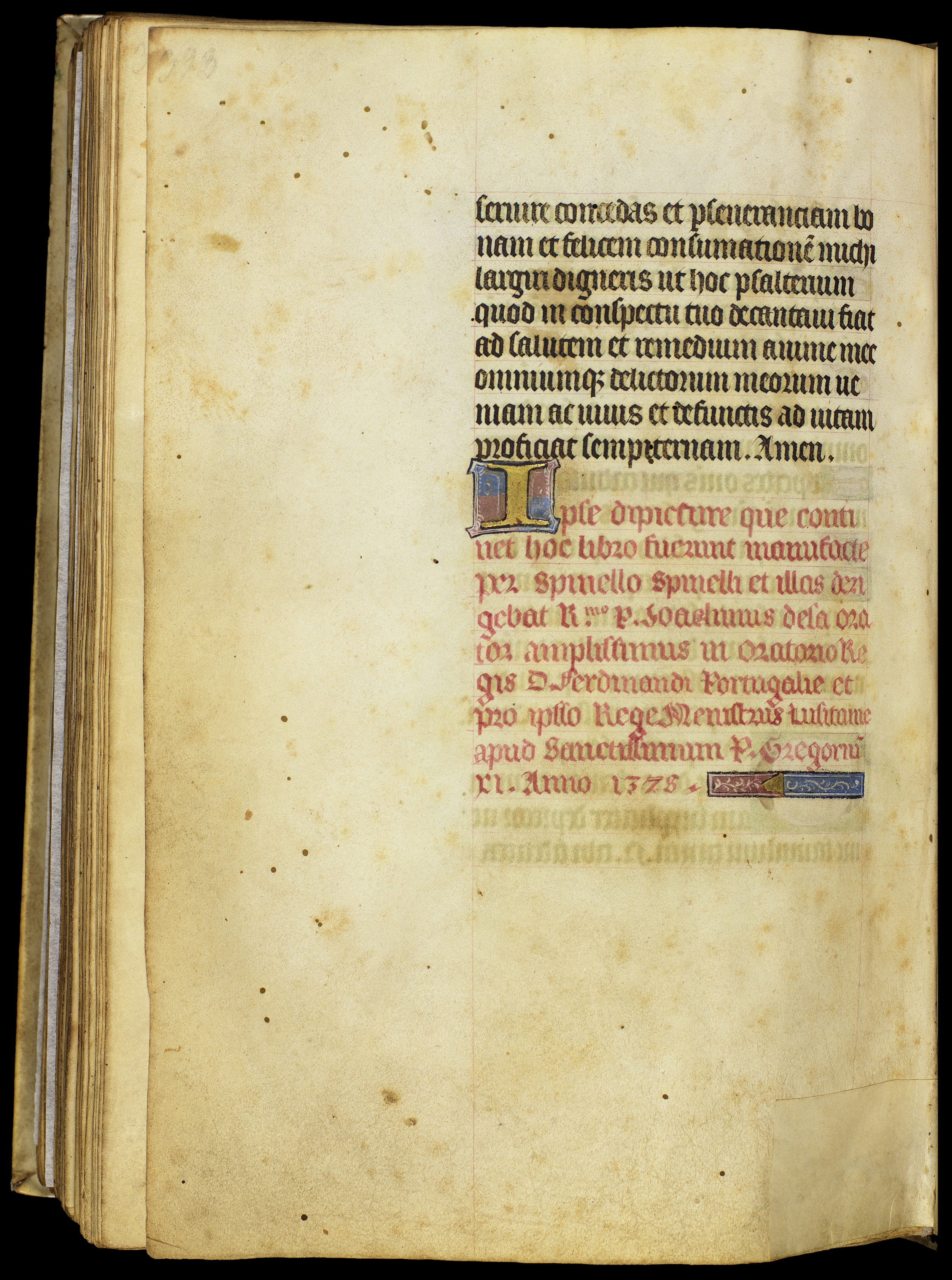
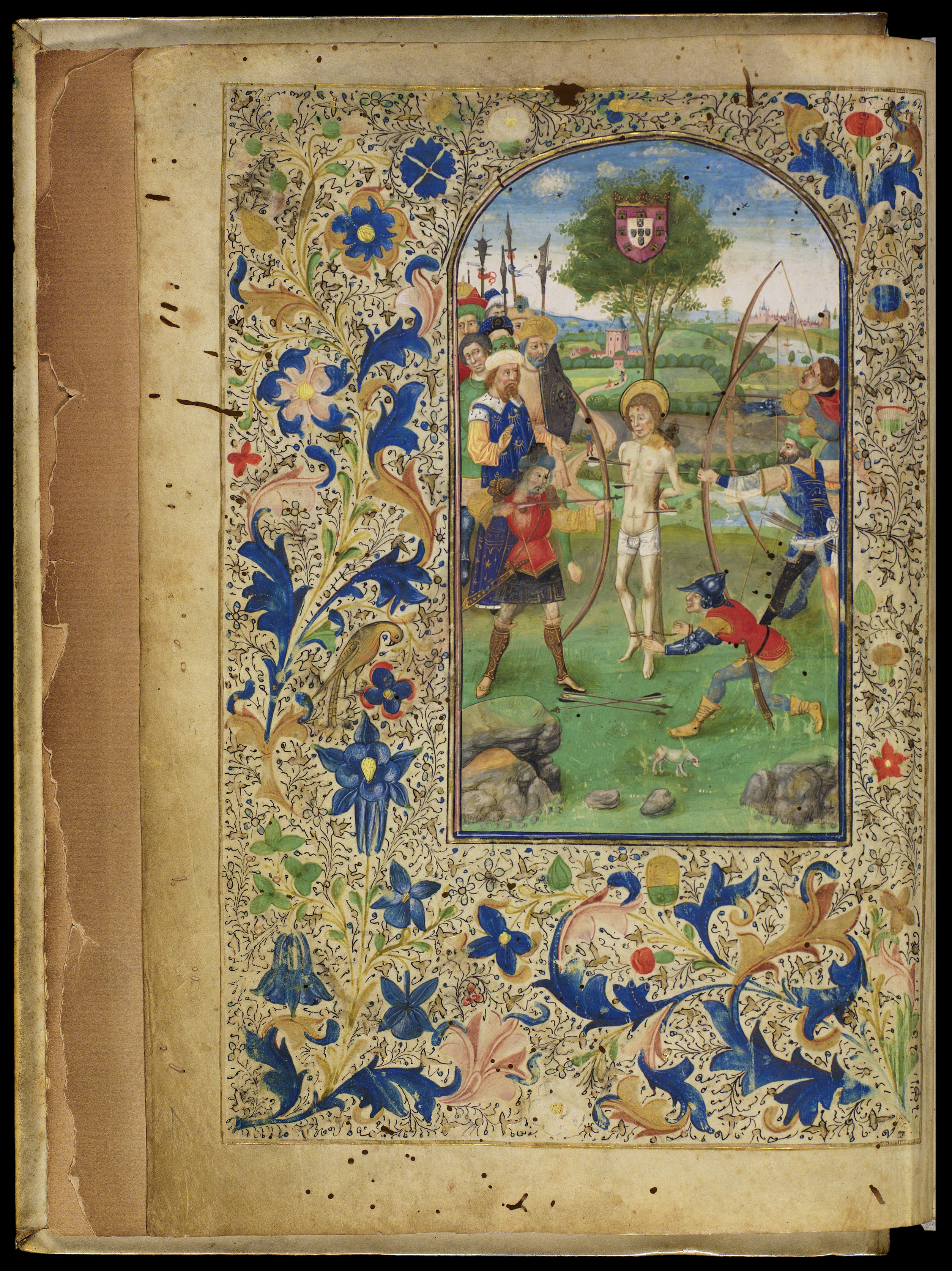
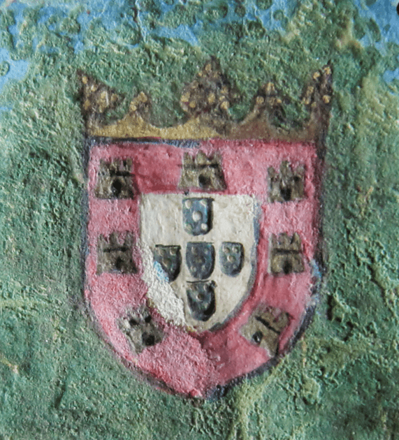
The Brazilian National Library (Biblioteca Nacional – BN) has a relevant collection of these prayer books from the 15\(^{th}\) century, including the luxurious 50,1,1 (call number CF-50,01,001), known as the “Book of Hours of D. Fernando”. It came from the Royal Library of Portugal to Rio de Janeiro shortly after the arrival of the Portuguese Royal Family, and for a long time, it was believed to have been illuminated by the Tuscan artist Spinello Spinelli for King Fernando I of Portugal (1345-1383) in the year 1378, due to the information in its colophon. The inscription, highly detailed, follows the final text on folio 199v (the last folio is blank), is written in Latin, in red gothic script, and decorated with an illuminated initial and a line filler, as shown in Figure 1. In addition, there is a coat of arms of the Portuguese crown on the opening folio (1v), which also contains a full-page miniature of the Martyrdom of Saint Sebastian, see Figure 2.
However, since the 1940s, all this narrative has been disputed by scholars through formal, stylistic, and textual analyses, raising issues of authenticity, origin, and the route of the codex (Berge, 1945; Berge, 1976; Fróes, 2011; Marrow, 2002). According to Marrow, 2002, this is an example from ca. 1460 to Sarum use (the liturgical Christian rite practiced in the region of Salisbury, England) and belongs to a rare subgroup of Flemish (probably from Bruges) books of hours produced for use in England in the third quarter of the 15\(^{th}\) century. All scholars agree that the colophon is an addition of untrue information much later than its production; the coat of arms would have been added at the same time and by the same hand. In addition, the originality of the illumination where the coat of arms is located was also called into question due, among other things, to its unusual position, appearing before the calendar
(Fróes, 2011).
.
The codex has 200 parchment folios measuring 242 \(\times\) 173 mm and contains a large number of illuminations, 32 of which are full-page or half-page, in addition to countless capitals with historic and ornamented details and other decorative elements.
Material characterization
The study presented in this paper came about with the goal of helping to elucidate the questions raised by scholars, cited above. The study specific goals, in its first phase, were to identify the color palette and characterize the pigments and materials of which the manuscript was made; identify differences between pigments, materials, and construction techniques to help correlate the obtained palette to a region, period, or workshop. In the future, we aim to add new techniques to help to identify underlying drawings and paintings, as well as fillers and binders.
To carry out the study, a multi-analytical and non-invasive approach was adopted, in accordance with the practices already established by several multidisciplinary teams and adapting them to the availability of equipment and resources (Aceto et al., 2012; Melo et al., 2011; Miliani et al., 2010).
To preserve the safety and integrity of the manuscript, non-invasive analysis and examination methods that could be performed in situ and carried out in the BN Restoration Laboratory were selected.
All equipment – except the photographic ones – belong to the IFRJ laboratories and were jointly operated by the professors and technicians of the Institute and by the conservator responsible for the project.
The data processing and analysis of results were also jointly carried out by the professors and the conservator.
The techniques and equipment used are briefly described below and will be referred to in this text by their most used acronyms.
Optical Microscopy (OM)
Used for observation and recording of details, colors, morphology, identification of construction techniques, and signs of degradation. Images were recorded with a digital camera integrated into an Olympus SZX16 stereomicroscope, with an SDF-PLAPO-1XPF objective and zoom lens, resulting in a total magnification of 7× to 115×, under normal and low light, with the aid of coaxial and ring illuminators.
X-Ray Fluorescence (XRF)
Elementary analysis technique through dispersion energies, acting in depth and allowing the identification of upper and lower layers. XRF measures were carried out with a TRACER IV portable system from Bruker, composed by an X-ray tube with Rh anode, which can operate at a maximum voltage of 40 kV and maximum current of 60 \(\mu\)A. The system was equipped with an X-ray detector, model XFlash® (10 mm\(^2\)) Silicon Drift Detector (SDD), thermoelectrically cooled at – 15°C, whose resolution for Mn-K\(\alpha\) energy is 145 eV and that can perform up to 10 kcps. XRF spectra were acquired during 40 seconds, placing the system at 5 millimeters from each point, with the tube operating at 40 kV and 10 \(\mu\)A. Spot beam size: ca. 5 mm in diameter. Thirteen parchment folios and a paper flyleaf were analyzed (on average, three points for each color), ranging from 3 points on the paper flyleaf to 62 points on folio 1v, resulting in a total of 190 points. Spectra were analyzed with Artax software.
Fiber Optic Reflectance Spectroscopy (FORS) in visible light (VIS)
Surface analysis technique based on reflection and absorption of light (visible, ultraviolet, and infrared). The Ocean Optics USB 4000 spectrometer was used, with a spectral range of 350 to 900 nanometers (nm), in the following set up and conditions: 12 scans, 5 boxcar, Illuminant D65, Observer 10 degrees; calibration on a standard black and a white Spectralon®. Application: optical fiber positioned at 45º from the sample with the aid of a probe positioned over the illuminations. The diameter of the analysis area is about 3 mm. The results were compared with those available in open access databases or from the team’s previous work (Cosentino, 2021). Spectra were visualized and recorded with the Ocean Optics Spectra Suite software. A number of 22 folios and a total of 176 points were analyzed.
Principal Components Analysis (PCA)
Chemometric technique in which the data matrix is decomposed into three other matrices (scores, weights, and error). The graphs of scores generated by the projection of PC2 \(\times\) PC1 and by PC3 \(\times\) PC1 show how much the samples are similar to each other and were used to evaluate possible groups of pigments from the FORS spectra. Calculations were performed using The Unscrambler® X 10 software (Camo Analytics).
Technical photography
Digital documentation of details of the manuscript in diffused, transmitted, raking, and ultraviolet light was carried out with a Canon T3i camera with an 18-55mm zoom lens. For ultraviolet fluorescence (UVF) photography, a standard black light lamp was used. The complete digital images were performed in 2008 with a Hasselblad H1 Ixpress 384 by the FBN Digitization Laboratory and are available online in compressed form in "", ().
The points analyzed by XRF and FORS were marked in a digital map for every folio with aid of the software Movida (v. 1).
Results and discussion
Characterization of pigments, materials and techniques
The results of the analytical techniques of XRF, FORS, and OM – with the aid of PCA – were combined to obtain a comprehensive characterization within the limits of these techniques. In this section, the results obtained are described: colorants, materials, and color construction techniques applied in the manuscript and, more specifically, in the coat of arms of f. 1v and in the colophon of f. 199v. The summary of the results obtained by the analytical techniques are shown in Table 1.
| Color / material | Category / hue | Present in the original body (O) or additions (A) | Pigment or material | Coloring substance / formula |
|---|---|---|---|---|
| writing inks | light and dark brown | O | iron gall ink | iron sulfate: Fe\(_2\)SO\(_4\) + Cu, Zn etc. |
| pink and carmine | O | brazilwood lake + lead white | brazilein | |
| carmine | A (199v) | red organic lake (madder/cochineal?) | alizarin and purpurin/ carminic acid | |
| white | inorganic | O/A (1v and 199v) | lead white | basic Pb carbonate: (PbCO\(_3\))\(_2\)·Pb(OH)\(_2\) |
| red | inorganic | O | vermillion (+ minium) | mercury sulfide: HgS |
| organic (pinks and carmines) | O | organic lake: brazilwood | brazilein | |
| A (199v and 1v) | organic lake (madder/cochineal?) | alizarin and purpurin/ carminic acid | ||
| orange | inorganic | O | minium | lead tetroxide: Pb\(_3\)O\(_4\) |
| yellow | inorganic | O | lead-tin yellow | lead tin oxide: Pb\(_2\)SnO\(_4\) (probably type I) |
| O | massicot (?) | lead oxide: PbO | ||
| gold | burnished | O/A (199v) | gold leaf | Au |
| matte | O / A (1v) | gold powder | Au | |
| ground | calcium-based | O | gypsum or calcite | calcium sulfate or calcium carbonate: CaSO\(_4\) / CaCO\(_3\) |
| iron-based | A (199v) | Armenian bole | iron oxide | |
| green | inorganic | O | malachite | Cu\(_2\) CO\(_3\) (OH) \(_2\) |
| blue | inorganic | O | azurite | Cu\(_3\) (CO\(_3\))\(_2\)(OH)\(_2\) |
| A (199v and, probably,1v) | lapis lazuli / ultramarine blue | lazurite (Na,Ca)\(_8\) [(S,Cl,SO\(_4\),OH)\(_2\) (Al\(_6\)Si\(_{6}\)O\(_{24}\))] | ||
| organic | O | (probably) indigo | indigotin | |
| silver | metal | O | shell silver (oxidized) | metallic silver: Ag / silver sulfide: Ag\(_2\) S |
Parchment
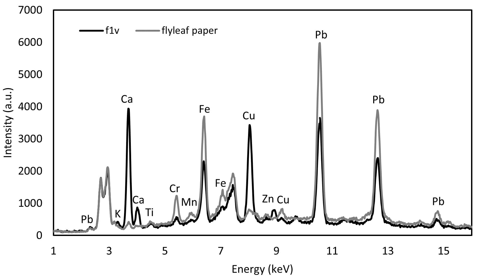
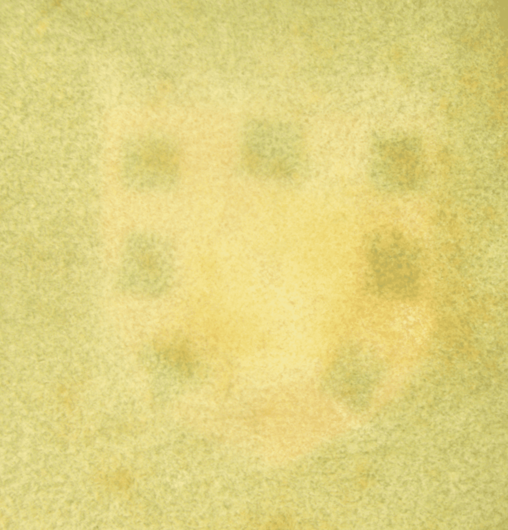
The support of the illuminations and the text, a thin and white parchment, was first analyzed by XRF in areas free of illumination or writing. In the thirteen selected folios, elements normally present in historical parchments were detected, such as calcium (Ca), iron (Fe), potassium (K), and sulfur (S) (Bicchieri et al., 2008; Guerra et al., 2013). In addition, copper (Cu) and lead (Pb) were detected in intense peaks, while titanium (Ti), manganese (Mn), zinc (Zn), and strontium (Sr) appeared in trace amounts. It is noteworthy, though, the presence of chromium (Cr) traces in the first three and the penultimate folios (folios 1v, 2r, 3r, and 199v). To investigate the origin of these elements, one of the flyleaves from the bookbinding was also analyzed, which is visible in Figure 2. The analysis of the paper, which is in direct contact with the first folio, showed Pb and Fe as major elements, in addition to considerable Cr peaks, while Ca peaks are low, as can be seen in Figure 3.
Thus, we considered the hypothesis of migration of Pb, Fe and Cr ions from the flyleaves to the first and final folios; on the other hand, Pb and Cu are found in considerable amounts in most of the folios analyzed. We also consider the possibility of the presence of a layer of lead white preparation on the parchment (de Viguerie et al., 2018; Watteeuw & Van Bos, 2008) and the migration of copper ions from the copper pigments (Coccato et al., 2017). Apart from these differences, all the analyzed folios presented similar results, with no indication of the addition of folios to the original body of the manuscript.
Writing inks
The inks used in the text writing – except the rubrics, which will be discussed later – were characterized by XRF as iron gall inks due to the presence of Fe, Cu, and Zn. The seven folios analyzed were classified into three groups according to the tonality of the brown hues, as shown in Table 2. It was observed that the darker inks contain a greater proportion of Fe, while the lighter ones contain more Cu; Zn, a minor element in the three groups, appears at a trace level in the lighter ones – which is in agreement with the findings of Hahn, 2010, also indicating the use of inks from different origins or recipes, and possibly, different hands (Aceto et al., 2017). Additionally, the FORS analysis confirmed the characterization of the inks as iron gall due to the increase in reflectance in the red range of the spectrum (Aceto et al., 2014).
| Images | Ink tone | Folios | Elementary composition (inks and parchment) |
|---|---|---|---|
 | light brown | 2r, 3r | Ca, Cu, Fe, Pb, (Zn, K, S, Ti, Mn) |
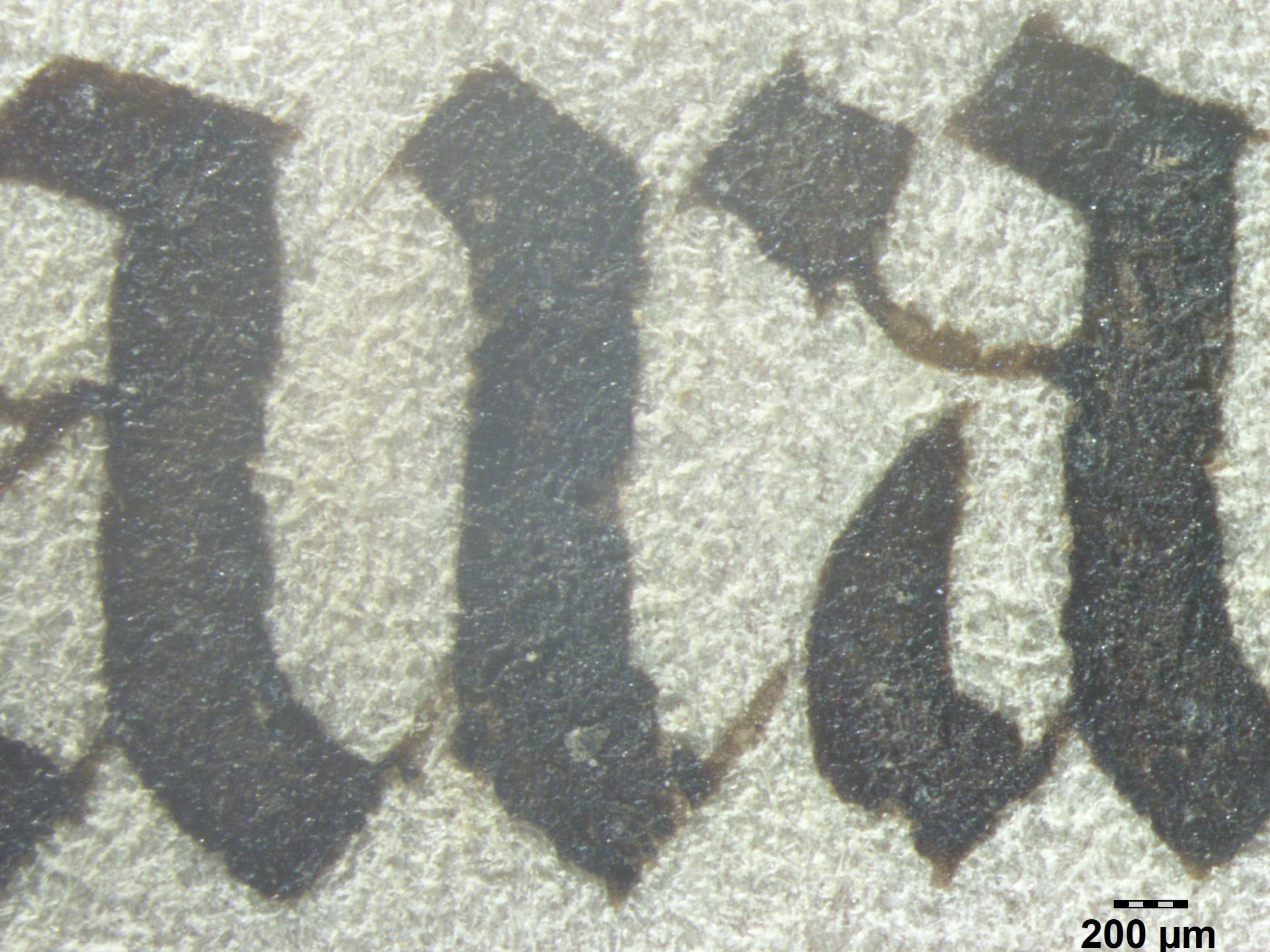 | dark brown | 11r | Fe, Cu, Ca, Pb, Zn, (K, S, Ti, Mn) |
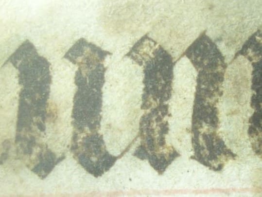 | dark brown | 194v 199v | Fe, Ca, Cu, Pb, Zn, (K, S, Ti, Mn) |
Whites
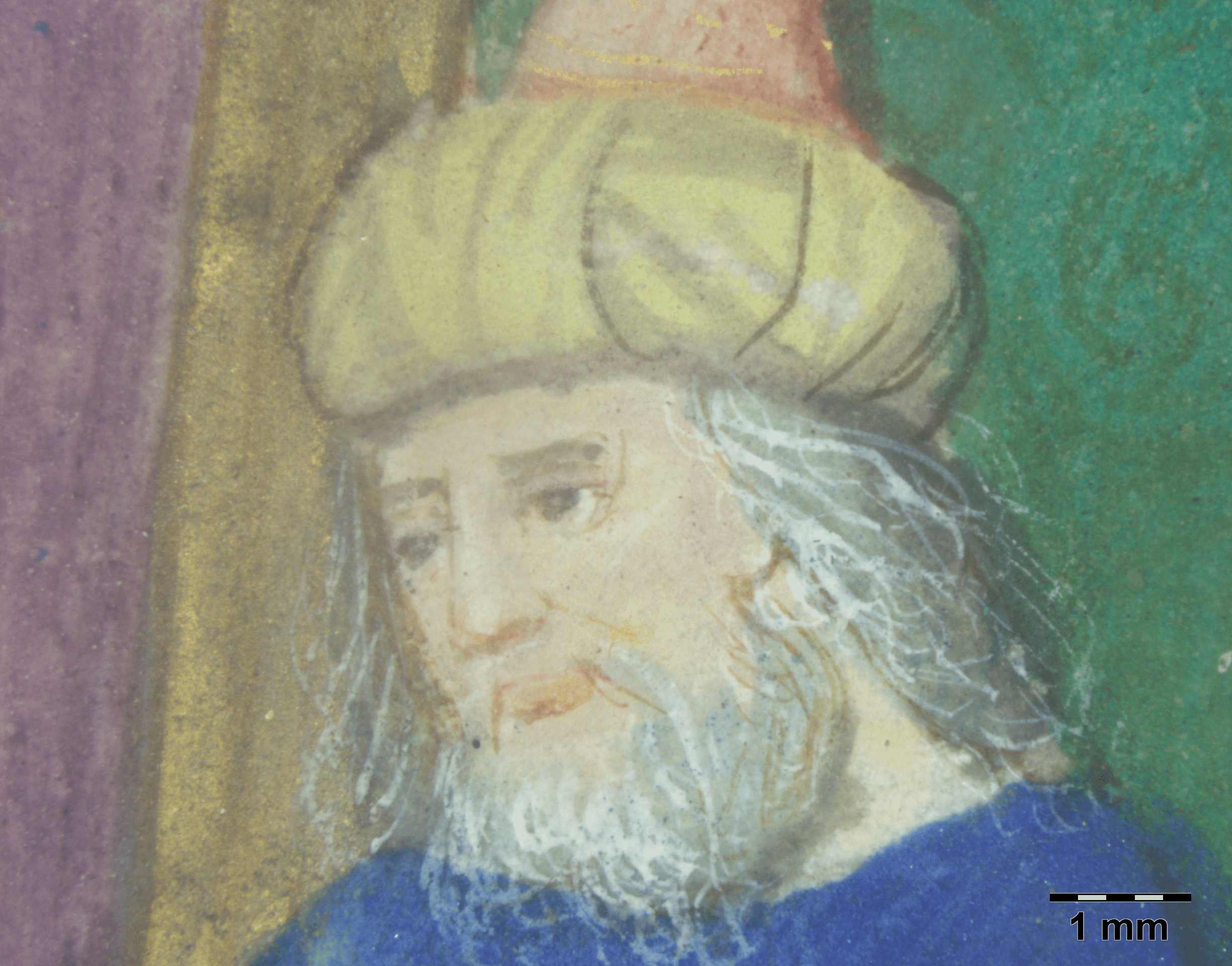
Lead white [(PbCO\(_3\))\(_2\)·Pb(OH)\(_2\)] was applied pure or mixed with other colors and as the base of flesh tones, Figure 4. It was identified by XRF by the intense Pb peaks. As mentioned before, lead white could also have been used as a thin layer of preparation on the parchment, as lead lines were detected on all folios analyzed, as shown in Figure 3.
Other whites such as calcite (calcium carbonate) and gypsum (calcium sulfate) may have been used as fillers for other pigments or as a mordant for lakes, indicated by intense Ca peaks; calcite was also used for the preparation of the parchment for writing and illuminating (Ricciardi & Beers, 2016).
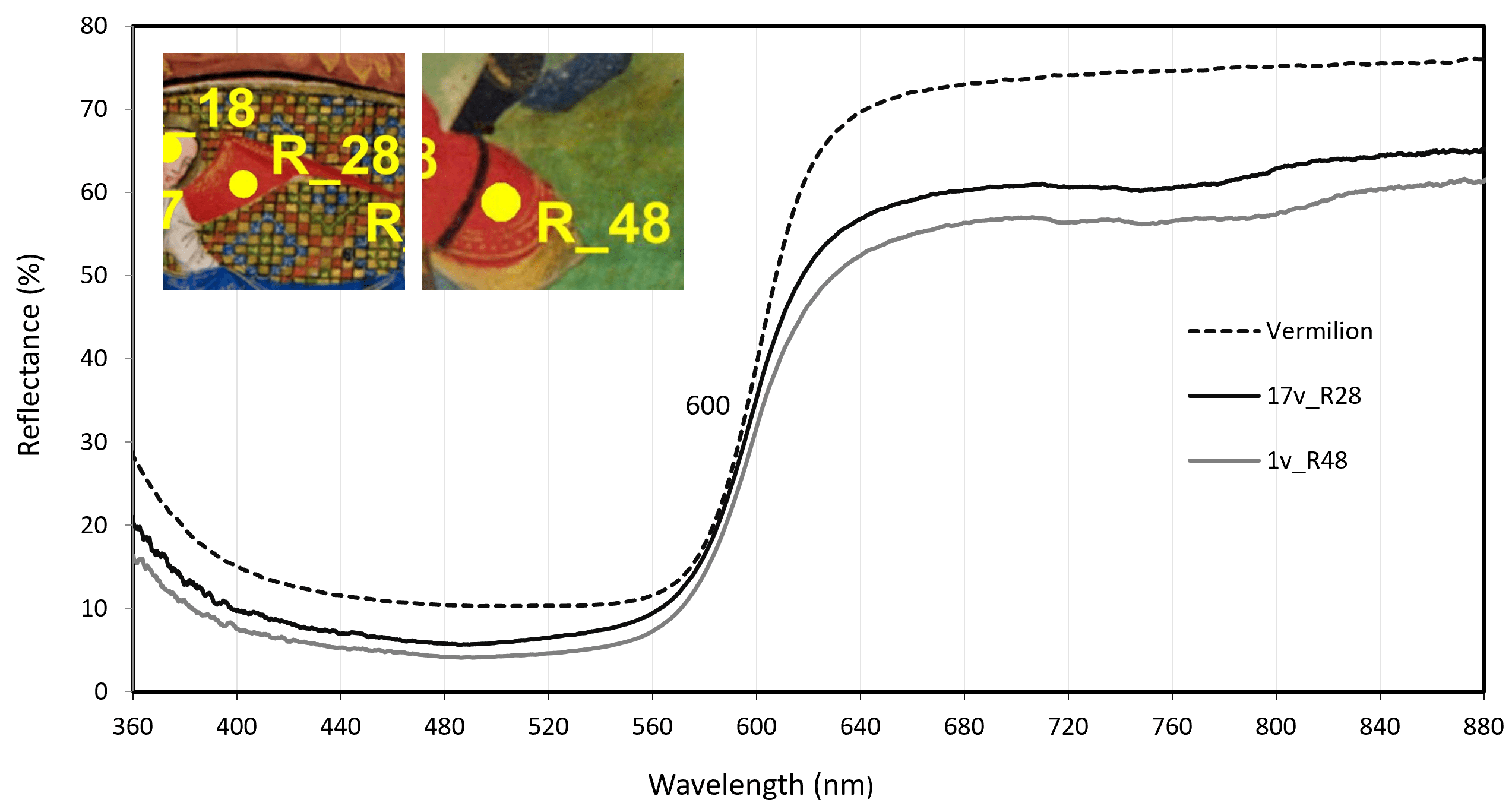
Reds and oranges
The XRF analyses of the red and orange hues mostly detected mercury (Hg) and lead (Pb), and also traces of sulfur (S), revealing the presence of the pigments vermilion (HgS) and minium or red lead (Pb\(_3\)O\(_4\)). Vermilion was easily confirmed by FORS - see Figure 5. Minium was not found pure, but admixed to vermilion, revealing that red and orange-red colors were constructed with different proportions of these pigments, as blends or overlaps, which may include lead-tin yellow (indicated by Pb and Sn) and an organic red (indicated by its translucency).
Greens
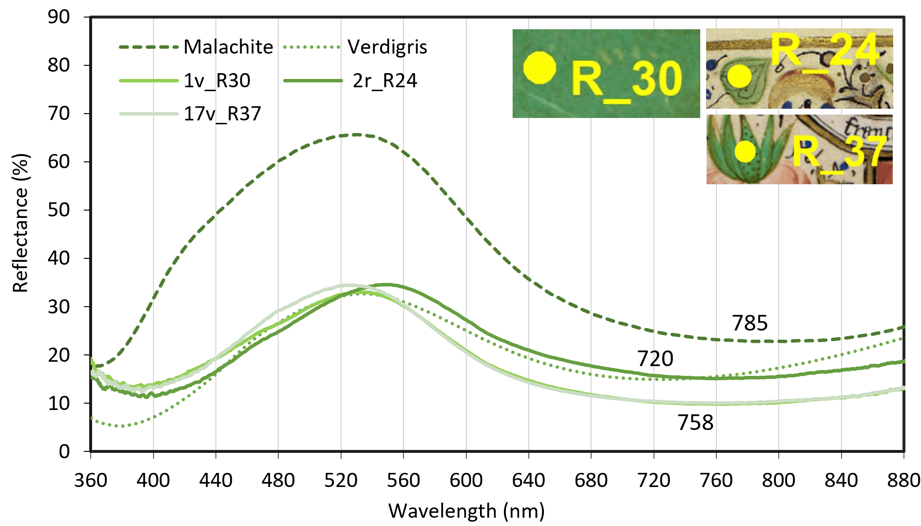
The element Cu was detected in green colors, indicating the presence of green copper pigments such as malachite or verdigris – which cannot be differentiated by XRF. With FORS, it was not possible to differentiate them clearly by their apparent absorbances maximum (a.a.m.) because the analyzed greens fall between the a.a.m. of the pigments references, although they are nearer the spectral features of malachite - see Figure 6.
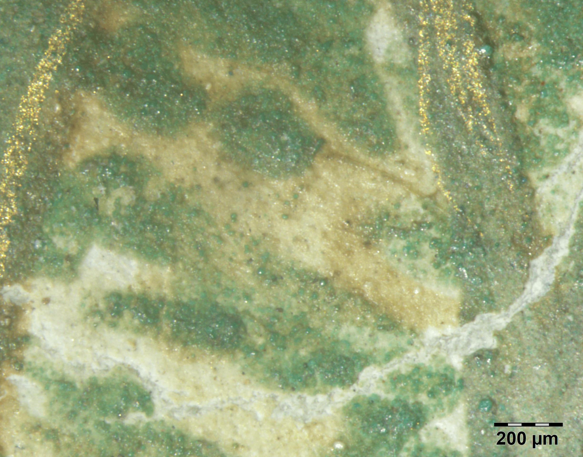
However, observation through OM of irregular grains, characteristic of the ground mineral, indicated the presence of malachite [Cu\(_2\)(CO\(_3\))(OH)\(_2\)] - see Figure 7.
Yellows
The light yellow applied purely in some details or as a component of brown tones was identified by XRF as lead-tin yellow (type I: Pb\(_2\)SnO\(_4\) or type II: PbSn\(_{1-x}\)Si\(_x\)O\(_3\)) due to the presence of the elements Pb and Sn.
However, at some points, the presence of Sn was unclear, while most FORS spectra indicated greater similarity with massicot (PbO) rather than with lead-tin yellow, Figure 8. Therefore, further investigation with the aid of complementary techniques is necessary to confirm this hypothesis.

Gold
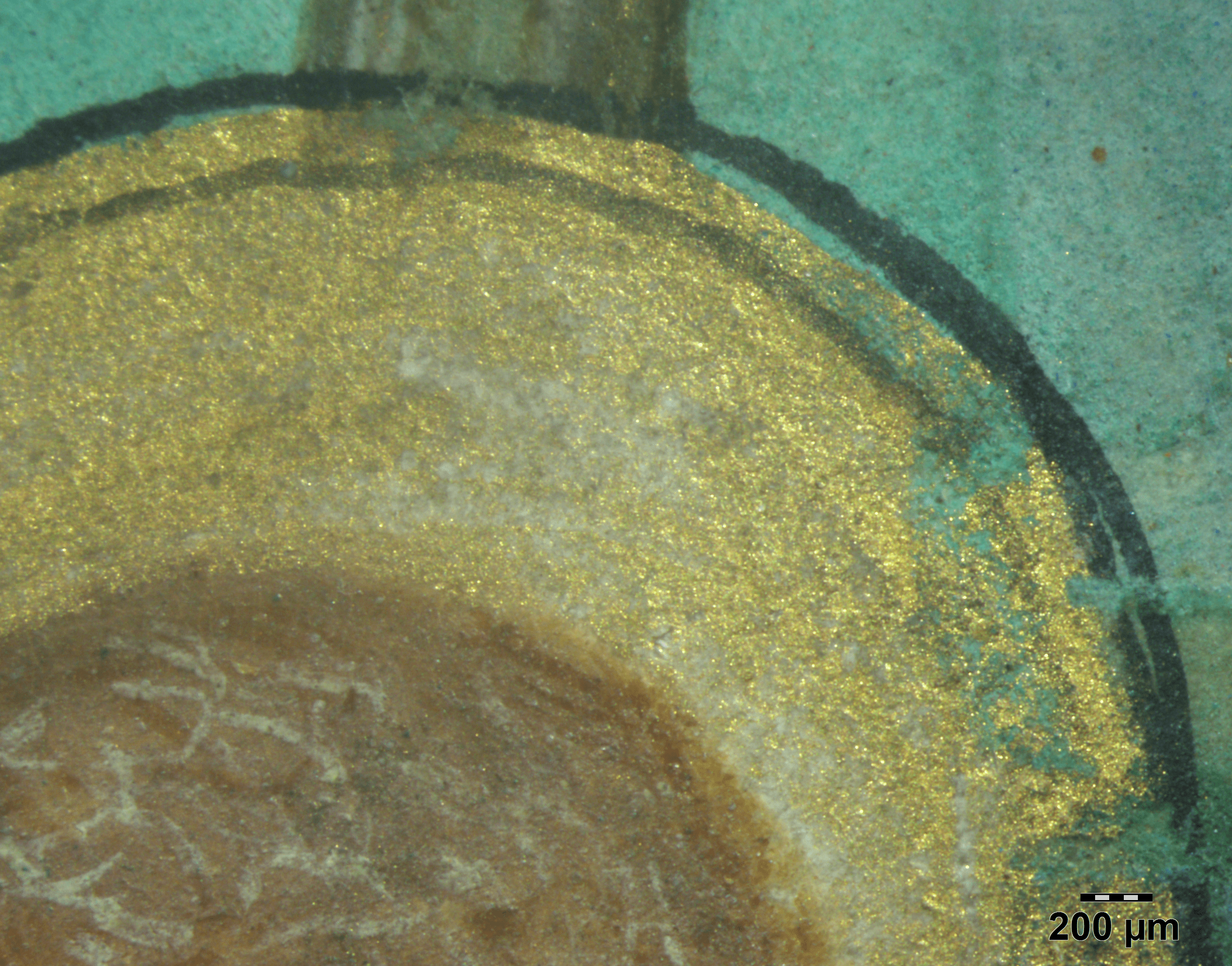
Gold was widely applied in this book of hours, both in leaf and powder form. This second technique was also called shell gold and has a matte appearance (as in flowers, acanthus leaves, garments, backgrounds, etc.). In the flat gilding technique, gold leaf was laid down directly on an area prepared with a transparent binding medium (as in saints halos). In the raised technique, a base of gesso is applied (as in initials, frames, ivy leaves, etc.) and a shine is given through burnishing (Brown, 2018). The element Au is easily detected by XRF, as well the calcium, which appear in intense peaks in the raised gilding of the analyzed set of folios original body, indicating the presence of a calcium-based preparation layer (gypsum or calcite), visible in some areas of wear of the gold leaf, Figure 9.
In the colophon of folio 199v, the gold leaf, applied on the initial “I” and the line filler, is flat and has a coarser appearance, with less shine. Some gaps in this foil allow a reddish background to be seen with the naked eye; XRF analysis detected, in addition to Au, Fe as a major element, indicating the presence of iron oxide – a component of the Armenian bole – in the preparation layer; and Ag in trace level, which indicates that the gold leaf contains silver in its alloy. In contrast, the XRF spectrum of gold foil from other initials shows Ca as the major element, indicating the presence of a calcium ground. A comparative is shown in Figure 10 (note that Fe, Cu and Zn peaks are also due to the script ink on the backside). Regarding the coat of arms of f. 1v, the presence of Au was confirmed by XRF, and OM images indicate that gold was applied in liquid form in the crown.


Blues
Blue is the most common color in this manuscript, applied in different saturations to acanthus leaves, flowers, skies, backgrounds of capital letters, garments, etc. In the typical palette of the period, the most common blue pigments available were the minerals azurite and lapis lazuli and the organic dye indigo.
Lapis lazuli was the pigment with the greatest economic and symbolic value, being generally reserved for the highest-ranking figures, such as the Virgin, and most used in luxury manuscripts – but, contrary to what was expected, this pigment was not detected in the original body of the manuscript.
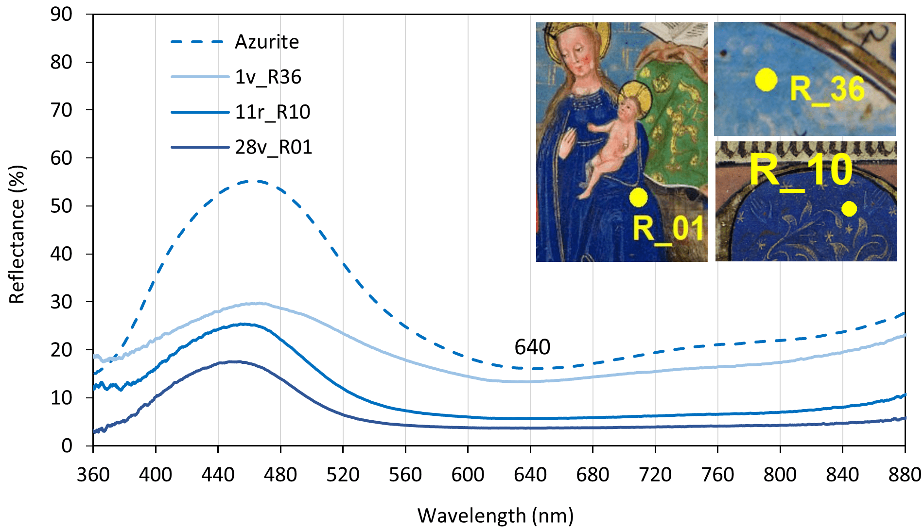
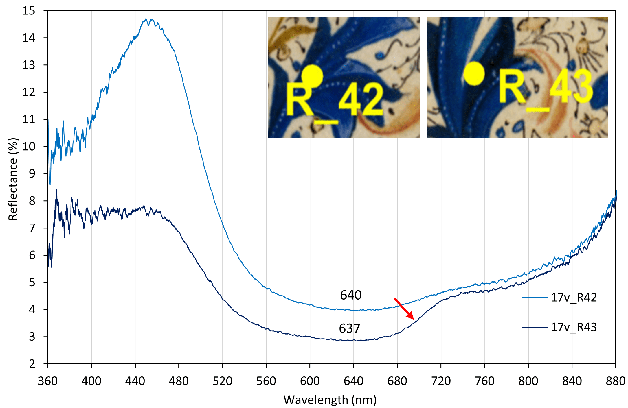
Both XRF and FORS analyses indicated only azurite [2CuCO\(_3\)·Cu(OH)\(_2\)], respectively due to the great intensity of Cu and the maximum absorption at 640 nm, Figure 11(a). The dark, translucent blue used in shadows is probably indigo, as the FORS spectrum suggests by maximum absorption around 660 nm, Figure 11(b).
(a)
(b)
However, when analyzing the blue background of the illuminated initial and the line filler of the colophon of f. 199v, lapis lazuli [Na\(_8\)[Al\(_6\)Si\(_6\)O\(_{24}\)]Sn] was identified by FORS – due to the maximum absorption close to 600 nm and the typical rise in reflectance in the red region -see Figure 12.
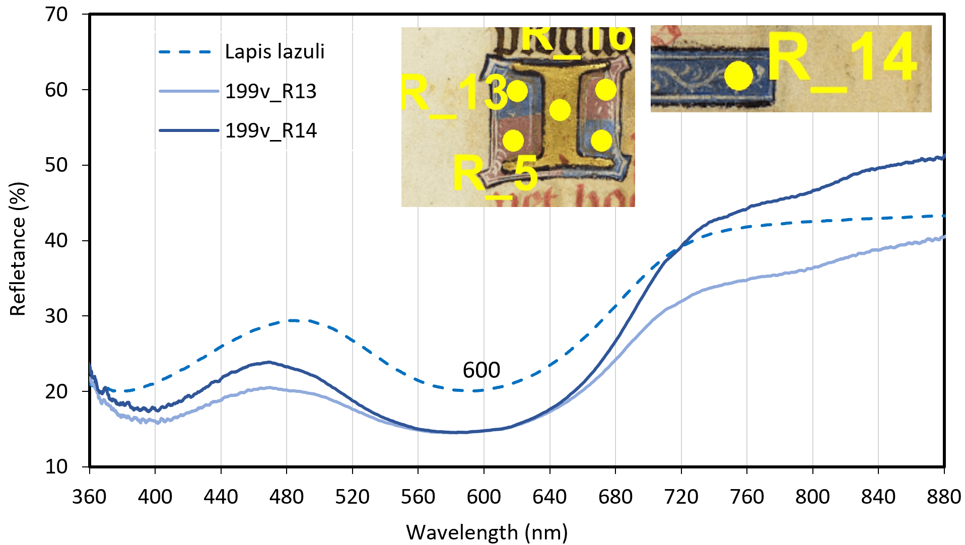
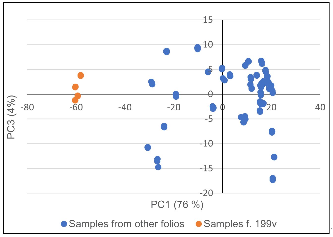
Besides portable XRF could not detect light lapis lazuli elements (Al and Si), this result was corroborated by the PCA chemometric calculations performed with FORS spectral data, revealing that the f. 199v blue points are different from all the others and similar to each other, Figure 13(a) and 13(b).
(a)
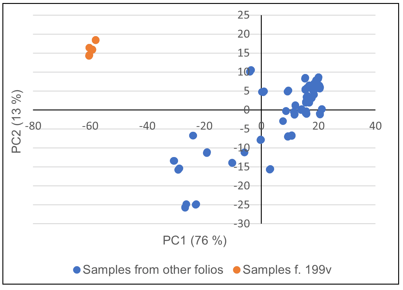
(b)
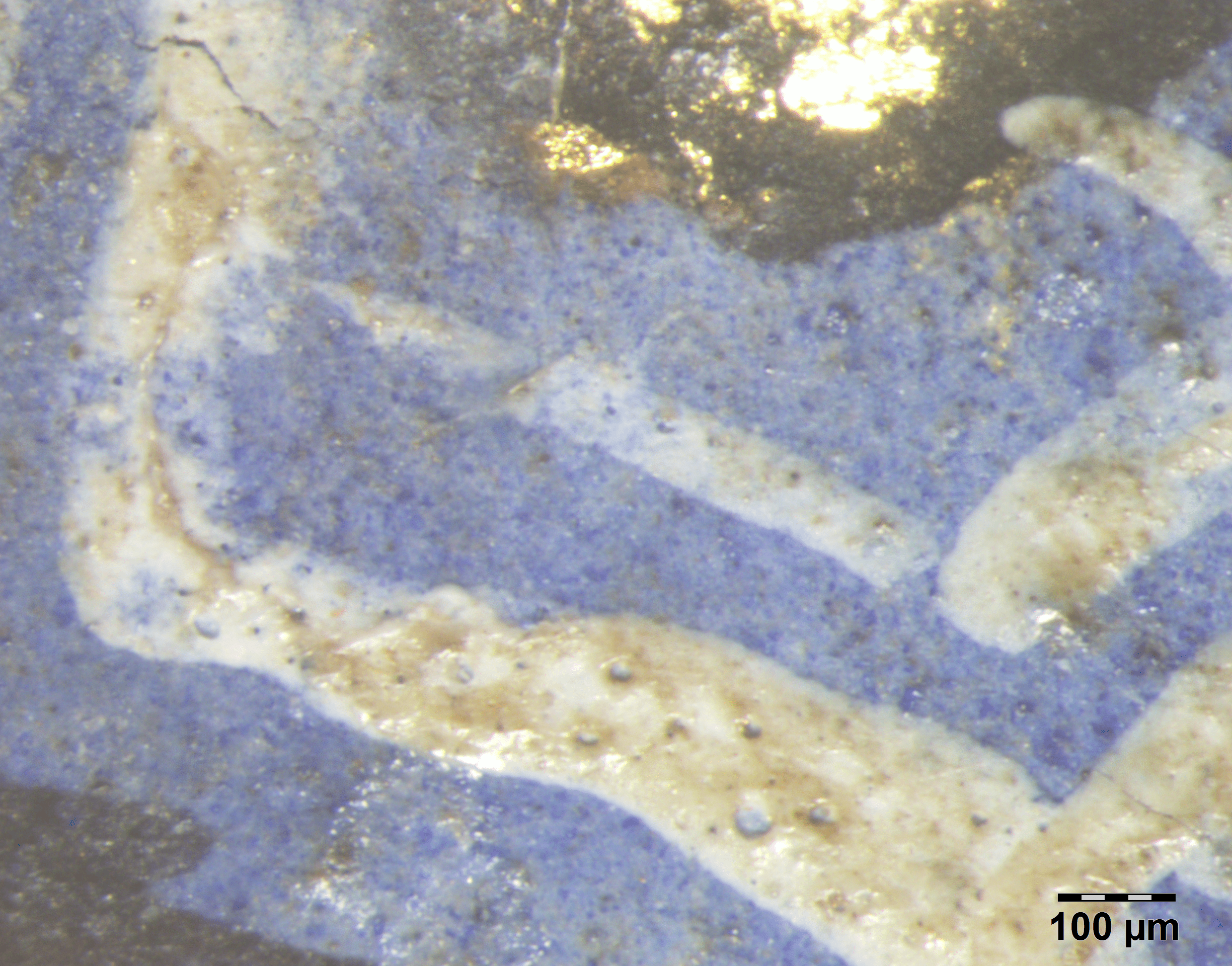
When observing the OM images, the differences between the blues become evident: high granulometry and intense blue hue in azurite, Figure 14(a), while lapis lazuli shows a pale violet-blue color with scattered larger and more intense blue grains, Figure 14(b) – probably from what is called ultramarine ash (the result of the last extractions of the pigment) mixed with lead white.
(a)
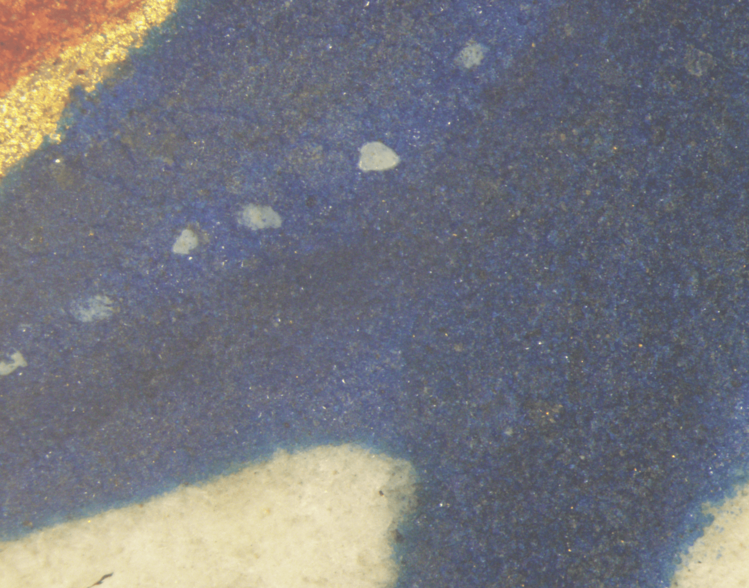
(b)
Pinks and carmines
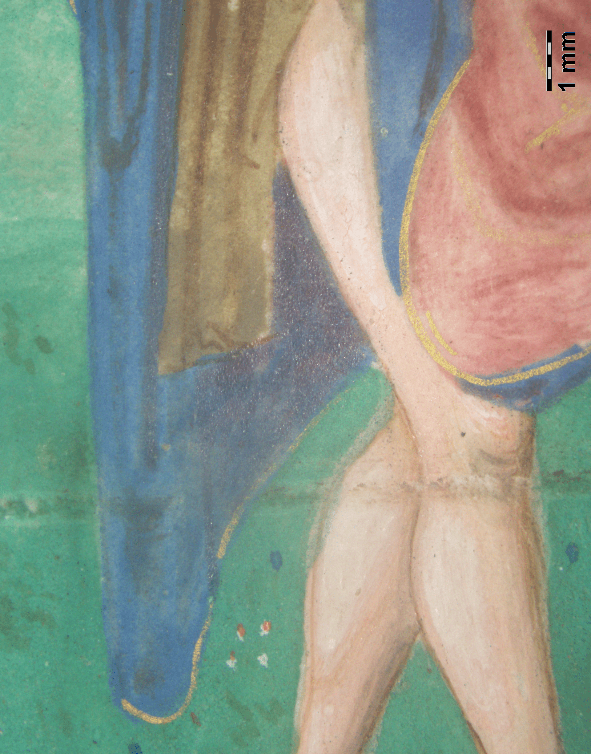
The various shades of pink and carmine red were extensively applied throughout the manuscript, whether in the rubrics, ruling or illuminations. The XRF analysis indicated its organic origin by excluding elements other than Ca, which may be part of the composition of the colorant as a filler (or from the parchment itself), and Pb, which indicates the mixture with lead white pigment in opaque pink and flesh tones - see Figure 15(a).
Among the red dyes available in the 15\(^{th}\) century, there were those derived from plants such as madder and brazilwood, or from insects, such as kermes and European cochineals – which were replaced by other species from the Americas since the 16\(^{th}\) century (Porter, 2006). The FORS technique is particularly useful in identifying these dyes, as it is non-invasive and very sensitive to them (Bisulca et al., 2008; Fonseca et al., 2019).
The results were divided into three groups, where:
A) For the pinks and carmines of the original body (illuminations and initials), e.g. the flesh tone of the figure and the rose tone of the garment, Figure 15(a), or the lake applied on the golden acanthus, Figure 15(b), the reflectance spectra show a maximum absorption band around 555 nm – close to the reference standard and according to the literature for brazilwood lake (Aceto et al., 2014) - see Figure 16;
B) For the colophon script of f. 199v, the analyses indicated that the carmine-colored ink is different from the inks of the original body rubrics, resembling the pattern of anthraquinone-based dyes, such as madder and cochineal. In the spectra of the analyzed points, two sub-bands of absorption are observed, next to the points of the cochineal lake reference - see Figure 17, and a little closer to those pointed out by Aceto et al., 2014 for madder lake, which makes them the main hypotheses to be investigated in a future work;
C) The pinks on the border of the coat of arms of f. 1v, the background of the initial “I” and the line filler of f. 199v, which have similar saturated and opaque hues, indicate mixing with lead white (Pb was detected by XRF at a high intensity).
(a)
(b)
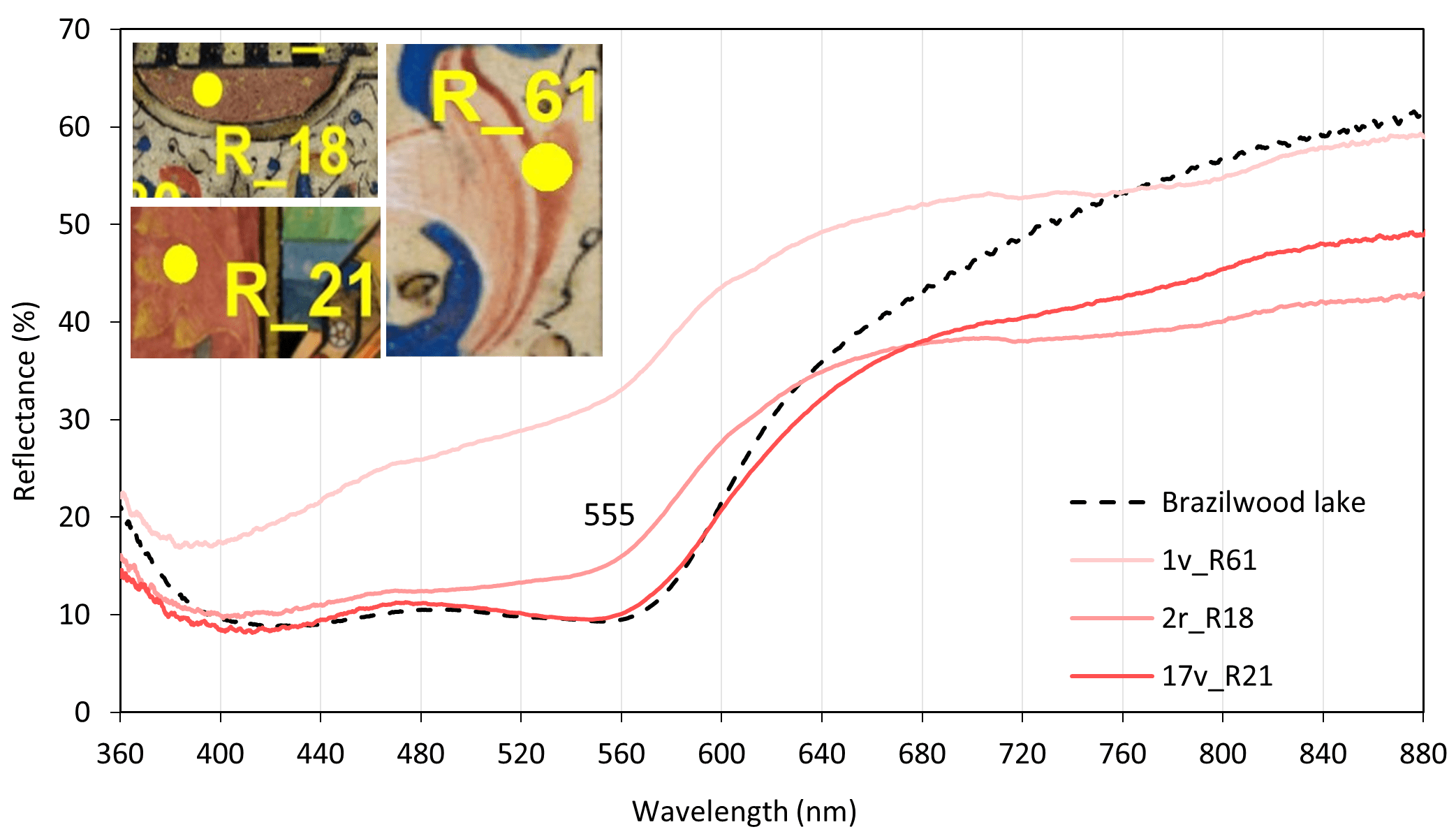

However, it was observed by FORS that the spectra of the first group differ a little from the spectra of the second group, Figure 18. Taking into account that, according to Bisulca et al., 2008, at high concentrations of red lakes on lead white, the absorption intensity is so great that only one broad band is observed instead of two sub-bands in the 500-560 nm range – and also (as it will be explained below) that this pink was painted on a first layer of vermilion, which could have had an influence – it is likely that the lake in these samples is the same as the one used to write the colophon.

Violets
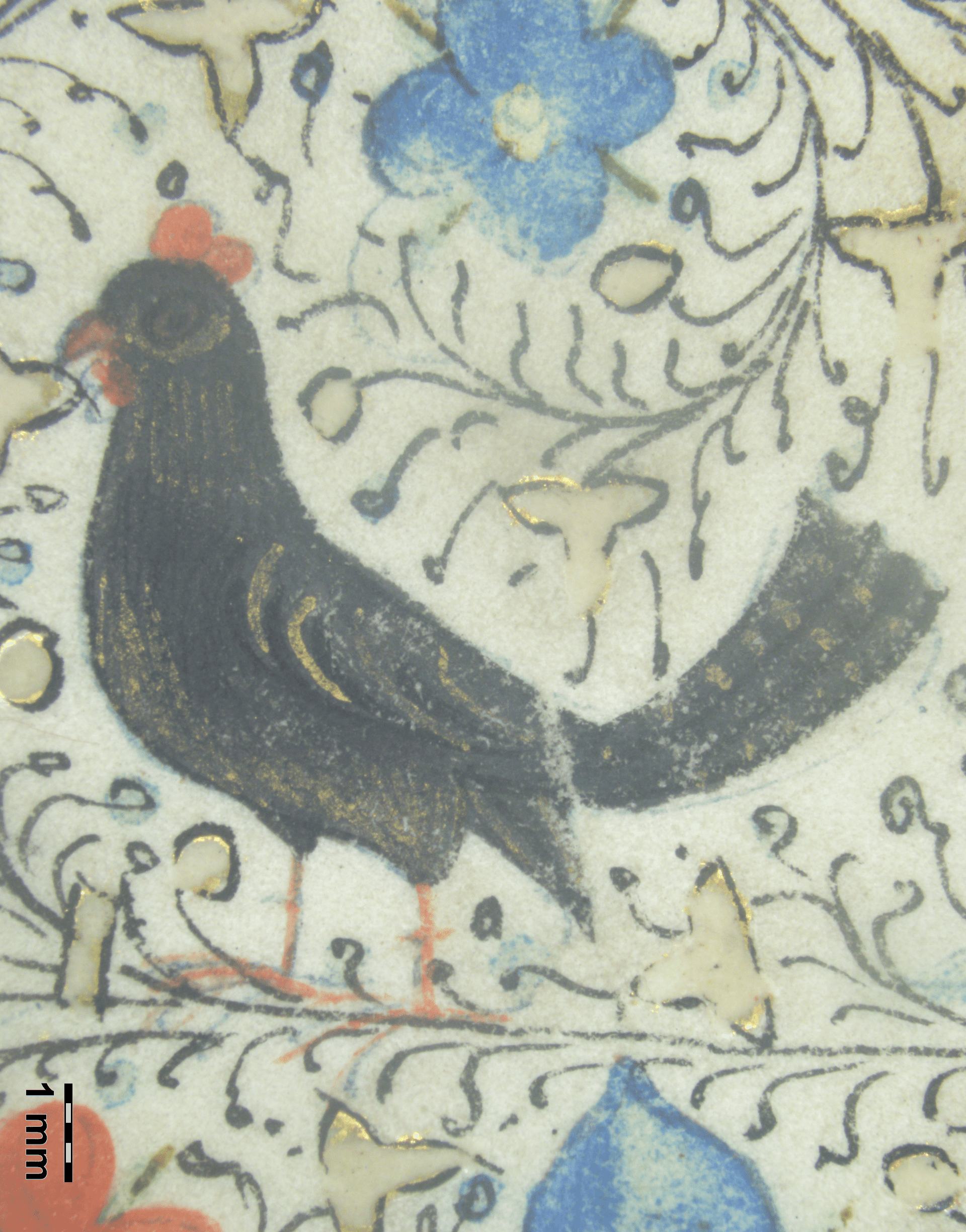
The violet color was applied to a few illuminations, particularly to Christ’s garments. According to Aceto et al., 2012, the identification of the components of this color is relatively difficult with only non-invasive techniques – however, despite the detection of Cu and Pb (ubiquitously present in the codex), both the microscopic observation (which has revealed no grains) and the FORS spectrum indicate the use of organic dyes, possibly a mixture of indigo with a red lake (Institute of Applied Physics, 2021). Examples are shown in Figure 19(a) (the violet zone of the background at left) and also an application of a red lake over a blue layer, as the violet-shaded area in the blue cloak of Figure 15(a).
(a)
(b)
Browns
These tones are present in the most diverse elements of the illuminations, such as hair, clothes, landscapes, animals, buildings, and shading, as in the figure’s outlines and background in Figure 19(a). They can be obtained with earth pigments or by mixing different pigments. XRF analyses detected Hg, Pb, Sn, and Cu, indicating the possibility of mixtures of vermilion, lead-tin yellow, and azurite or malachite; Au was also found in some of them, indicating a mixture of gold powder – evidencing the diversity in using pigment mixtures instead of earth pigments, which was a characteristic of northern European painting, as cited by de Viguerie et al., 2018.
Grays
Although it is likely that some grays contain dark pigments such as brown earths, carbon blacks and indigo, only grays with mixtures of lead white and azurite have been identified, as indicated by the presence of Cu and Pb by XRF and by visible blue grains under OM. Examples of gray shades and a gray beard are in Figure 19(a) and Figure 20(a).
(a)
(b)
Blacks
The black pigment, used mainly in outlines and drawing lines, such as foliage, Figure 19(b), can not be detected by the techniques used; according to the literature, it is likely, though, that a carbon-based pigment has been applied, such as lamp or coal black (Panayotova & Ricciardi, 2016).
Silver
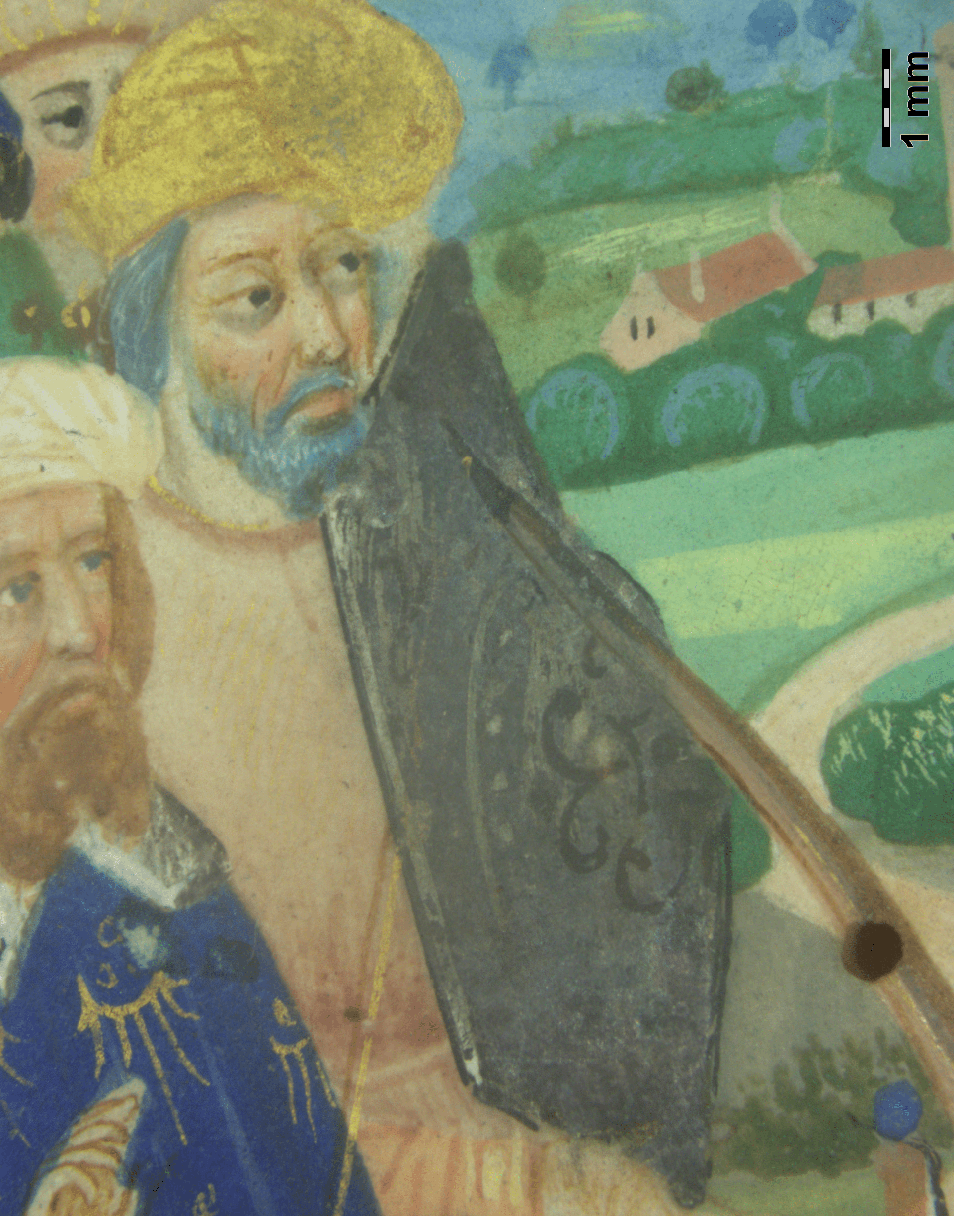
In this manuscript, silver, detected by XRF, was extensively applied in liquid form: in the representation of the metallic shine of weapons and armor, Figure 20(a), of light entering through windows, and in details of frames, Figure 20(b) – however, it is tarnished in most areas due to an oxidation process, probably forming silver sulfide (Ag\(_2\)S) (Araújo et al., 2018).
Results for specific questions
Illumination of f. 1v
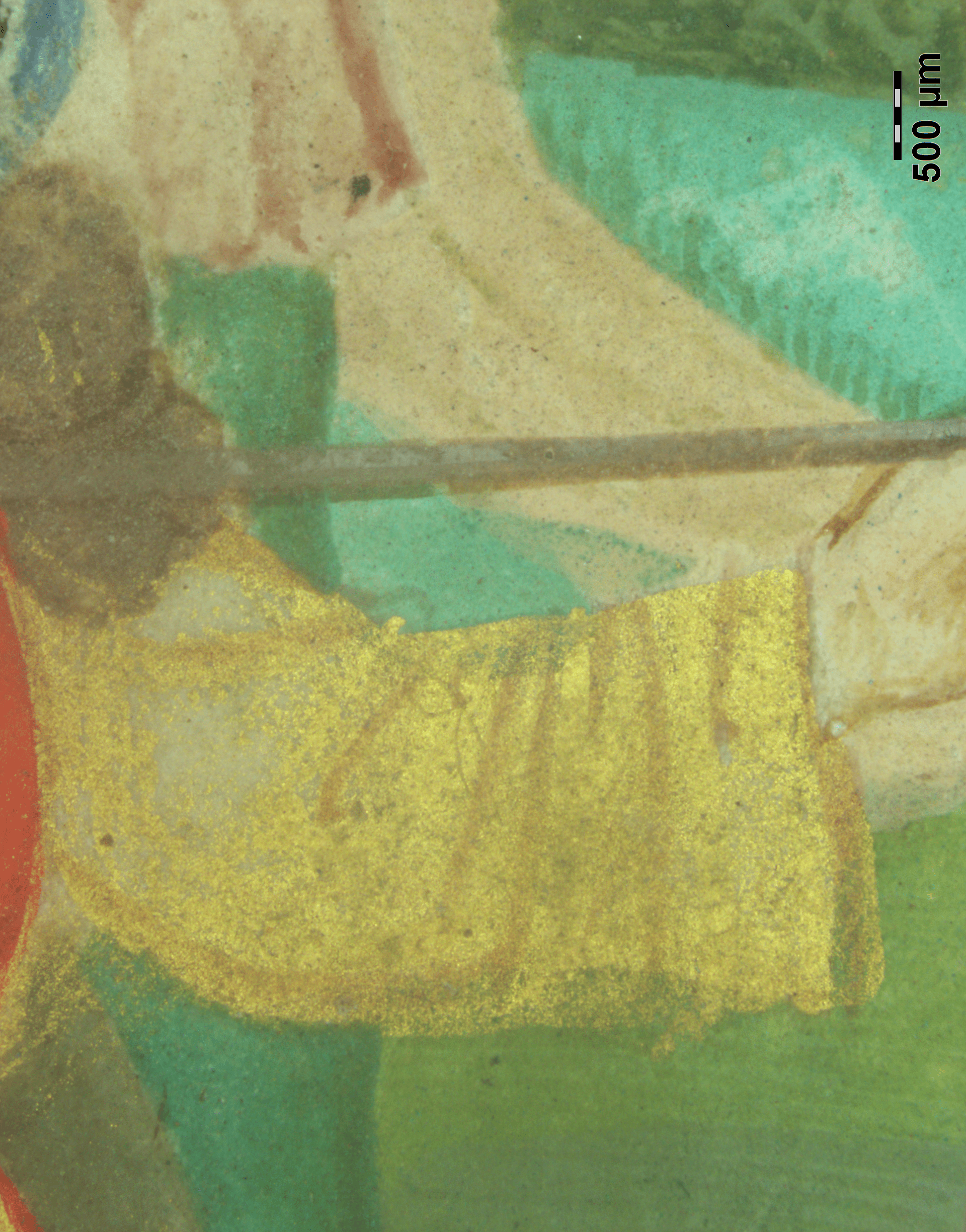
No different pigments were identified in the illumination of this folio in relation to other illuminations, but some differences were observed in the way the colors were applied, such as the use of gold: in addition to the border elements, it was applied as a pigment directly on the parchment in the S. Sebastian’s halo, Figure 21(a), and in the clothes of the soldiers and of the group of noble men on the left, Figure 21(b) – which was observed only in this folio. Furthermore, some formal aspects of the drawing composition and quire formation (which are out of the scope of this paper) differ from the rest of the manuscript. These differences indicate that, possibly, this illumination was not made in the same period or by the same workshop of illuminators.
(a)
(b)
The coat of arms of f. 1v
Due to the small dimensions of the elements of the coat of arms and the resolution limitations of the XRF equipment, the results in this region were difficult to evaluate. However, in general, they have indicated the following attributions: the element Ca, which can indicate an additive of the border pink lake, as well as a component of the parchment; Pb, indicative of white lead; Au, from the golden crown or the underlying layer; Ag, revealing the presence of silver, visible as tiny spots on the castles; Hg and S, indicatives of the pigment vermilion, and Cu, indicative of the presence of azurite, both from the underlying layer. The castles seem to be painted with a Cu-alloy, given the dark spots on the reverse side, Figure 22(a).
(a)
(b)
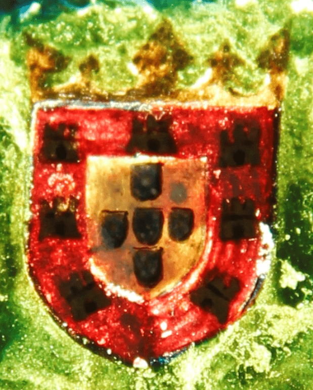
The same problem occurred with FORS analysis, both due to the resolution of the optical fiber and the difficulty of positioning the accessory precisely on the area to be analyzed. Besides this, it was possible to obtain a reasonable signal for the pink border, as commented in “Pinks and Carmines”. On the other hand, imaging tests were more effective. The photography with transmitted light allowed the visualization of an underlying painting layer, from which the presence of a quarterly coat of arms is inferred, being also perceptible the presence of some elements within these divisions, Figure 22(b).
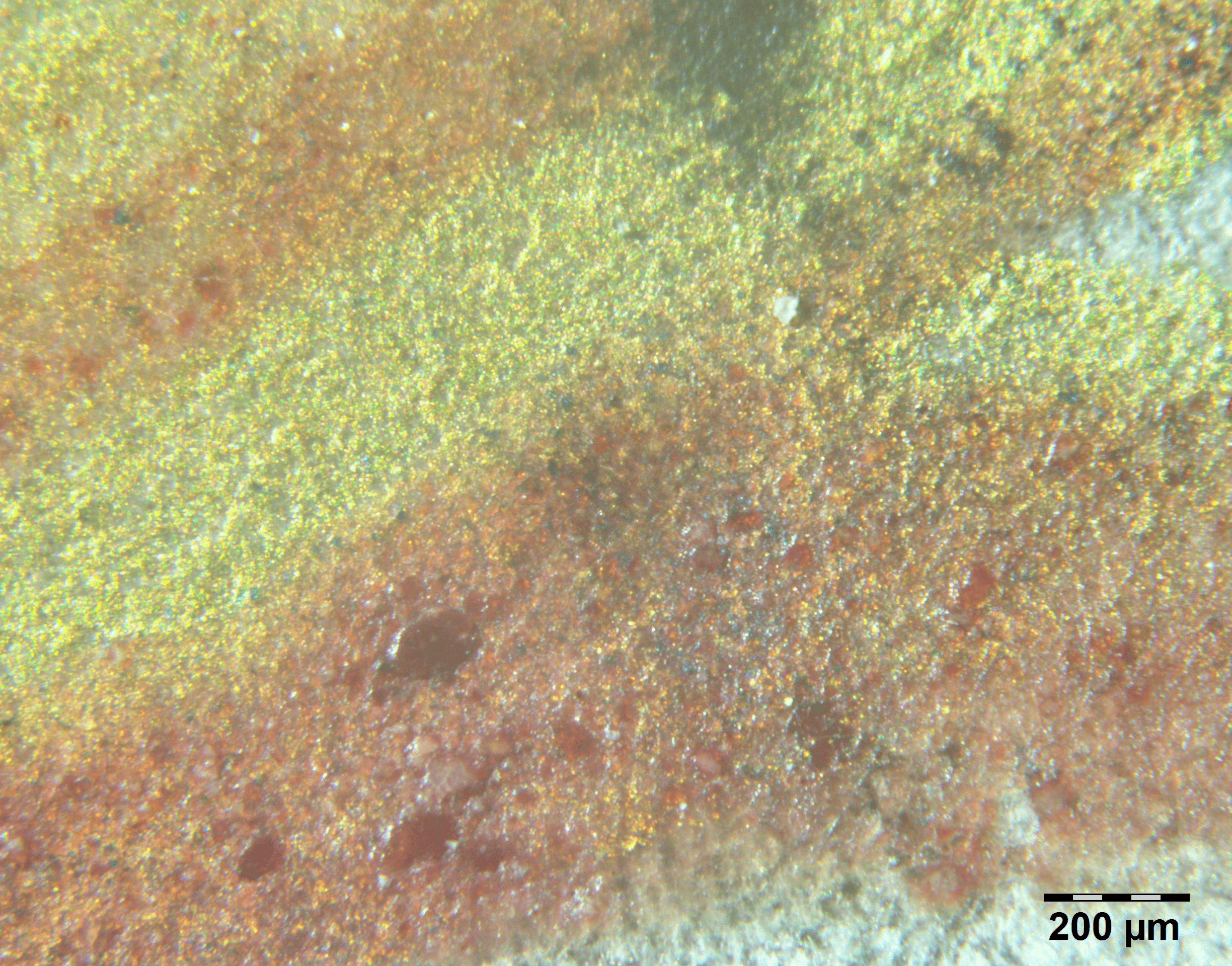
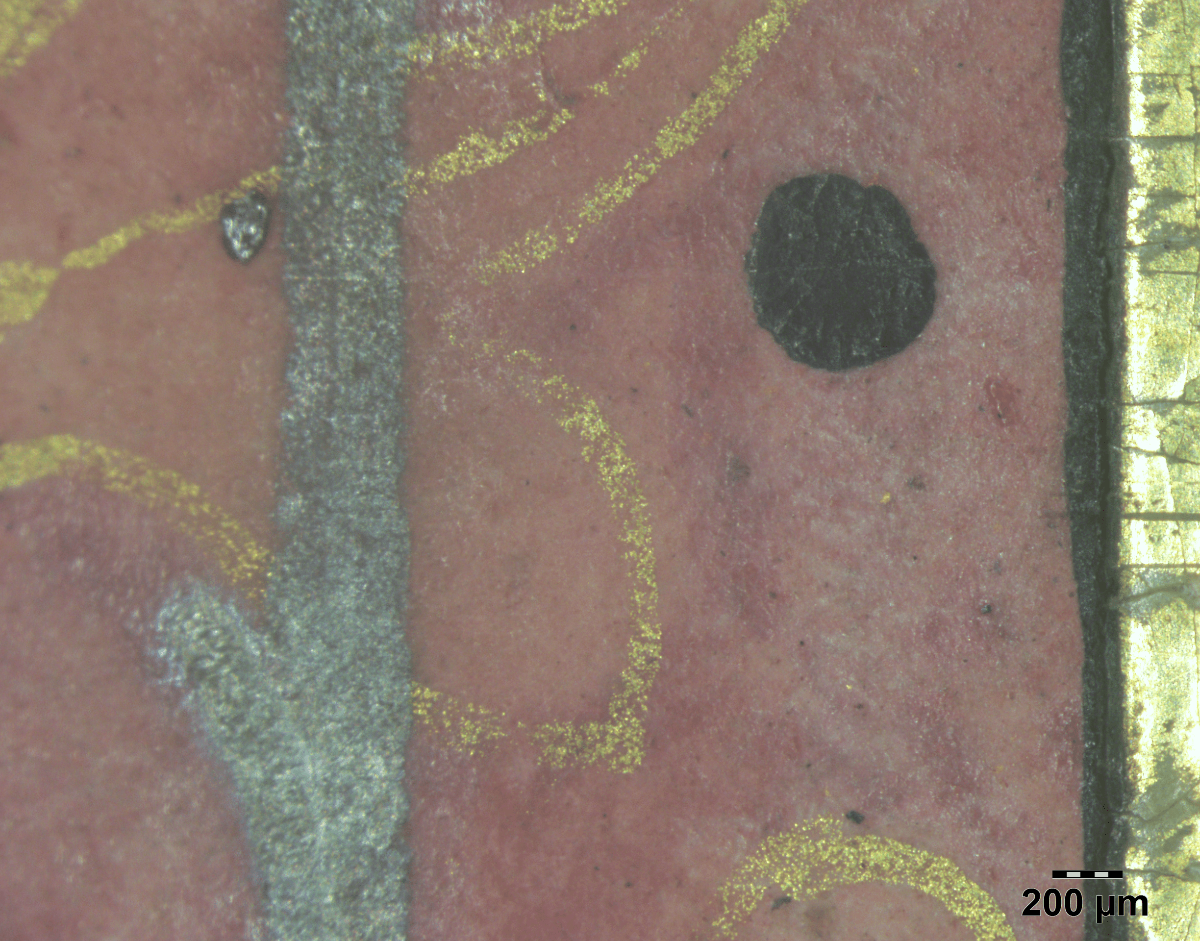
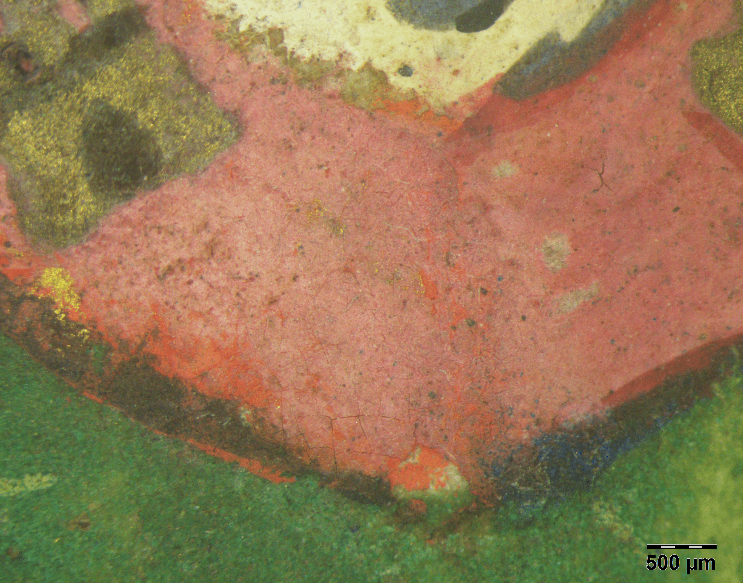
The photomicrographs, in turn, allowed a better observation of the layers of color and the losses of pigment and textures, specially with raking light, Figure 23(a). It was also possible to see the dispersed grains of pigments, identifying the presence of a layer of pink paint over a red one that occupies a lower quadrant and an opposite upper quadrant, and of a layer with blue pigment grains (visible in some spots) in the inverse quadrants as well, Figure 23(b). However, it seems that part of these pigments was scraped off. Therefore, further examinations and studies are needed for more accurate identification.
(a)
(b)
Conclusions
This study aimed to produce the most comprehensive characterization possible of the materials and techniques applied in the manuscript with the available set of techniques. In this phase of execution, paint additives and binders were not investigated. Although the techniques and examinations carried out so far have only allowed a partial characterization of colorants, they have allowed us to advance in the knowledge of this codex.
In the original body, lead white, vermilion, minium, lead-tin yellow, azurite, malachite, indigo and a brazilwood lake were identified or indicated; among metallic pigments, gold and silver, in addition to iron gall inks with different proportions of iron, copper and zinc; calcium, as a component of lake pigments and of the preparation of gilding. There is also the possibility that a second yellow pigment, massicot was applied.
In the colophon of folio 199v, lapis lazuli, white lead, and a red lake, possibly madder or cochineal, were found, in addition to gold leaf with little burnishing over a ground of Armenian bole. Therefore, about 50% of the palette was identified, about 35% have indications to be confirmed and the remaining 15% were not possible to investigate.
It was verified, thus, that the palette of the original body of the codex is varied, with intense and bright colors and with a great variety of tones and mixtures, as well as diverse color construction techniques – and, therefore, consistent with a typical palette of the northern Europe of the second half of the 15\(^{th}\) century – except for the absence of lapis lazuli, which would be expected specially given the high level of the illuminations. However, this pigment appears only in the illumination of the colophon which, together with the other discoveries mentioned, was confirmed as a later addition. The opaque pink color of the colophon illuminations, in turn, proved to be very similar to the same color as the border of the Portuguese Royal coat of arms, which is consistent with the hypothesis that both were made at the same time and by the same person. In addition to the presence of white lead, silver, and gold, some elements of pigments in the underlying layer were also detected in the coat of arms, identifying the presence of vermilion and azurite – revealing the existence of another coat of arms under the Portuguese shield, confirmed by preliminary examination of transmitted light and normal light microscopy.
On the other hand, no different pigments from the rest of the manuscript were found in the illumination of folio 1v, but rather the application of different techniques in the use of gold – which means that there is the possibility that it is not from the same set of illuminations. We were thus able to confirm some of the proposals: the inscription of the colophon on the existing folio 199v, as well as the Portuguese royal coat of arms on folio 1v, were in fact made after the writing of the manuscript; the codex was most likely originally made in the second half of the fifteenth century – as proposed by scholars as Berge, 1945, (1976) and Marrow, 2002. This demonstrates the validity of the use of instrumental analyses in an interdisciplinary approach to give pieces of evidence and help to elucidate issues related to the history of this remarkable illuminated manuscript.
Authors contributions
I.L.C. designed and wrote the paper, and preparared the images, graphs, and tables as well; R.P.F. coordinated and performed the XRF analyses; H.C.A.F. coordinated the FORS analyses; A.L.Q.B. performed the PCA analyses; A.L.O. performed the XRF measurements; L.S.P. and L.M.A. performed the FORS measurements; I.L.C. and each member of the team performed the respective interpretation of results. The OM setup was done by R.P.F. The photomicrographs and the technical photography were done by I.L.C. with aid of L.M.A. All members have read and approved the final text.
Conflicts of interest
The authors declare no conflict of interest.
Acknowledgments
We express our thanks to all the Biblioteca Nacional professionals involved in this project, namely the heads of the Manuscripts Section, Luciane Medeiros, and of the Restoration Laboratory, Jandira Flaeschen; and to the Preservation Coordinator, Jayme Spinelli Jr., for the arrangement of a Technical Cooperation Term with the Instituto Federal de Ciência e Tecnologia do Rio de Janeiro (IFRJ) – through which this interdisciplinary work was possible.
References
Aceto, M., Agostino, A., Fenoglio, G., Gulmini, M. B., & Pellizzi, E. (2012). Non invasive analysis of miniature paintings: Proposal for an analytical protocol. Spectrochimica Acta-Part A: Molecu-larand Biomolecular Spectroscopy, 91, 352–359. https://doi.org/10.1016/j.saa.2012.01.011
Aceto, M., Agostino, A., Fenoglio, G., Idone, A., Gul-mini, M., Picollo,M., Ricciardi, P., & Delaney, J. K.(2014).Characterisation of colourants on illuminated manuscripts by portable fibre optic UV-visible-NIR reflectance spectrophotometry. Analytical Methods, 6(5), 1488–1500. https://doi.org/10.1039/C3AY41951G
Aceto, M., Agostino, A., Fenoglio, G., Capra, V., Demaria, E., & Cancian, P. G. (2017). Characterisationof the different hands in the composition of a 14th century breviary by means of portable XRF analysis and complementary techniques. X-Ray Spectrometry, 46(4), 259–270.https://doi.org/10.1002/xrs.2752
Araújo, R., Nabais, P., Pombo Cardoso, I., Casanova, C., Lemos, A., & Melo, M. J. (2018). Silver paints in medieval manuscripts: A first molecular survey into their degradation. Heritage Science, 6(1), 1–13. https://doi.org/10.1186/s40494-018-0172-7
Berge, D. (1945). Um Livro de Horas do século XIV na Biblioteca Nacional. 2(1), 49-99.
Berge, D. (1976). Livros de Horas Manuscritos iluminados da Biblioteca Nacional do Rio de Janeiro. Fundação Biblioteca Nacional.
Bicchieri, Marina, Monti, Marco, Piantanida, Giovanna, Sodo, Armida & Tanasi, Maria Teresa (2008). Inside the parchment. In Proceedings [Proceedings]. 9th International Conference on NDT of Art, Jerusalem, Israel.
Bisulca, Christian, Picollo, Marcello, Bacci, Mauro & Kunzelman, Deborah (2008). Uv-Vis-Nir Reflectance Spectroscopy of Red Lakes in Paintings. In Proceedings [Proceedings]. 9th International Conference on NDT of Art, Jerusalem, Israel.
Brown, Michelle P. (2018). Understanding Illuminated Manuscripts: A Guide to Technical Terms. J. Paul Getty Museum.
Carvalho, I. L., Freitas, Ricardo P., Araújo, Herculano, Baddini, Amanda L., Oliveira, Ana L. C., Alves, Luana M. & Paula, L. (2019). Questões sobre o Livro de Horas dito de D. Fernando - estudo interdisciplinar sobre um códice iluminado da Biblioteca Nacional do Rio de Janeiro. In Instituto de Estudos Medievais, Conference.
Carvalho, I. L., Freitas, Ricardo P., Araújo, Herculano, Baddini, Amanda L., Paula, L., Oliveira, Ana L. C. & Alves, Luana M. (2020). Confirmação de elementos não originais por meio da caracterização da paleta do Livro de Horas CF-50,1,1 da Biblioteca Nacional. In M. A. Rizzutto, T. A. B. C. Sanjad, & F. O. Palácios (Eds.), Caderno de resumos expandidos 2 [Anais]. ANTECIPA 2020: Encontro da Associação Nacional de Pesquisa em Tecnologia e Ciência do Patrimônio, São Paulo, Brasil.
Coccato, A., Moens, L., & Vandenabeele, P. (2017). On the stability of mediaeval inorganic pigments: A literaturere view of the effect of climate, material selection, biological activity, analysis and conservation treatments. Heritage Science, 5(1), 29. https://doi.org/10.1186/s40494-017-0143-8
Cosentino, Antonio (2021). Download all FORS Spectra in a ZIP file: Reflectance Spectroscopy (350-950 nm) (Gorgias) Pigments-Checker Database. https://chsopensource.org/fors/
de Viguerie, L., Rochut, S., Alfed, M., Walter, P., Astier, S., Gontero, V., & Boulc’h, F. (2018). XRF and reflectance hyperspectral imaging on a15th-century illuminated manuscript: Combining imaging and quantitative analysis to understand the artist’s technique. Heritage Science, 6(1), 2–13. https://doi.org/10.1186/s40494-018-0185-9
Fonseca, B., Patterson, C. S., Ganio, M., MacLennan, D. & Trentelman, K. (2019). Seeing red: towards an improved protocol for the identification of madder- and cochineal-based pigments by fiber optics reflectance spectroscopy (FORS). 7(1), 1--15. https://doi.org/10.1186/s40494-019-0282-2
Fróes, V. L. (2011). O livro de horas dito de D.Fernando–Maravilha para ver e rezar.Anais da Biblioteca Nacional, 129, 85–135.
Guerra, M., Manso, M., Pessanha, S., LeGac, A., Longelin, S., Guilherme, A., Gil, M., Seruya, A. I., & Carvalho, M. L. (2013). X-Ray Fluorescence Spectrometry as a Diagnostic Tool in Characterization and Conservation of Manueline Illuminated Manuscripts. In P. Frediani, M. Frediani, & L. Rosi (Eds.), Cultural heritage: Protection, developments and international perspectives (pp.235–256). Nova Science Publishers.
Hahn, Oliver (2010). Analyses of iron gall and carbon inks by means of X-ray fluorescence analysis: A non-destructive approach in the field of archaeometry and conservation science. 31(1), 41--64.
Institute of Applied Physics. (2021).Fiber optics reflectance spectra (FORS) of pictorial material sinthe 270–1700 nm range. https://spectradb.ifac.cnr.it/
Livro de horas, uso de Sarum. (1450/1460). Manuscrito. http://acervo.bndigital.bn.br/sophia/index.asp?codigo_sophia=15259
Marrow, J. H. (2002). The Pembroke Psalter Hours. In B. Cardon, J. Van Der Stock, & D. Vanwijnsberghe (Eds.), “Als ich kan”: Liber amicorum in memory of Professor Dr. Maurits Smeyers (pp.882–883). Peeters.
Melo, M. J., Miranda, M. A. C., Lemos, A., Lopes, J. A. & Gonçalves, A. P. (2011). The Colour of Medieval Portuguese Illumination: an Interdisciplinary Approach. 152-173.
Miliani, C., Rosi, F., Brunetti, B. G. & Sgamellotti, A. (2010). In situ noninvasive study of artworks: The MOLAB multi-technique approach. 43(6), 728-738.
Panayotova, S., & Ricciardi, P. (2016). Masters’ Secrets. In S. Panayotova (Ed.), Colour: The Art & Science of Illuminated Manuscripts (pp.118–161). Harvey Miller Publishers.
Porter, C. (2006). The Use of Organic Colours in Manuscripts and the Importance of Correct Identification and Treatment Options. In B. Reissland (Ed.),Tintas y pigmentos.jornadas técnicas sobre restauración de documentos (pp.35–55). Gobierno de Navarra.
Ricciardi, P., & Beers, K. R. (2016). The illuminators’ palette. In S. Panayotova (Ed.), Colour: The Art & Science of Illuminated Manuscripts (pp.27–39). Harvey Miller Publishers.
Watteeuw, L. & Van Bos, M. (2008). The conservation assessment of an illuminated Book of Hours. Understanding craftsmanship through interdisciplinary research: preliminary investigation. In International Council of Museums, ICOM Committee for Conservation [Proceedings]. 15th Triennial Meeting, New Delhi, India..